 Indietro
The Cell Cycle: Mechanisms and Regulation
Guida di studio - Note intelligenti
 Appunti personalizzati basati sui tuoi materiali, ampliati con definizioni chiave, esempi e contesto.
Chapter 12: The Cell Cycle
Introduction to the Cell Cycle
The cell cycle is the series of events that take place in a cell leading to its division and duplication. It is fundamental to the continuity of life, ensuring that genetic information is faithfully transmitted from one generation to the next. Cell division is essential for reproduction, growth, development, and tissue repair in living organisms.
Cell Division: The process by which a parent cell divides into two or more daughter cells.
Types of Cell Division:
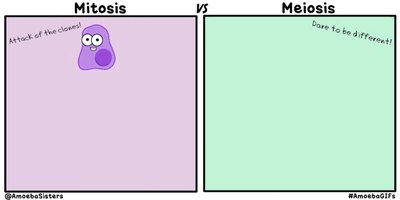
Mitosis: Maintains chromosome number; daughter cells are genetically identical to the parent cell.
Meiosis: Reduces chromosome number by half; produces gametes (sperm and eggs), leading to genetic variation.
Functions of Cell Division
Cell division serves different purposes in unicellular and multicellular organisms:
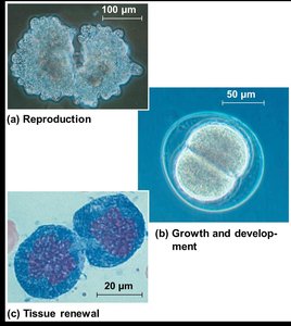
Unicellular Organisms: Cell division is a form of reproduction.
Multicellular Organisms:
Development from a fertilized cell
Growth
Repair of tissues
Organization of Genetic Material
Genome and Chromosomes
The genome is the complete set of genetic material in a cell. In prokaryotes, the genome is typically a single DNA molecule, while in eukaryotes, it consists of multiple DNA molecules organized into chromosomes. Eukaryotic cells also contain mitochondrial DNA (mtDNA) and, in plants, chloroplast DNA (cpDNA).
Chromosomes: Structures that package and organize DNA with proteins (chromatin).
Somatic Cells: Non-reproductive cells with two sets of chromosomes (diploid).
Gametes: Reproductive cells with one set of chromosomes (haploid).
Chromosome Number and Ploidy
Each species has a characteristic number of chromosomes. The number of chromosome sets is referred to as ploidy:
Diploid (2n): Two sets of chromosomes (e.g., humans: 46 chromosomes).
Haploid (n): One set of chromosomes (e.g., human gametes: 23 chromosomes).
Polyploid: More than two sets of chromosomes, common in plants.
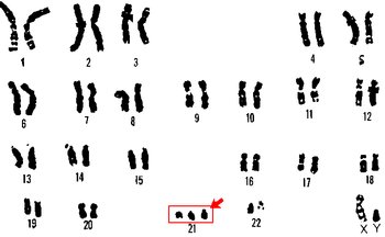
Abnormal chromosome numbers can lead to disorders such as Down Syndrome (trisomy 21).
Chromosome Structure and Replication
Before cell division, DNA is replicated, resulting in duplicated chromosomes composed of two sister chromatids joined at a region called the centromere. During division, sister chromatids separate, ensuring each daughter cell receives an identical set of chromosomes.

The Cell Cycle Phases
Overview of the Cell Cycle
The cell cycle consists of interphase (about 90% of the cycle) and the mitotic (M) phase. Interphase is subdivided into:
G1 phase (First Gap): Cell growth and normal metabolism.
S phase (Synthesis): DNA replication.
G2 phase (Second Gap): Preparation for cell division.
The M phase includes mitosis (nuclear division) and cytokinesis (cytoplasmic division).
Mitosis: Stages and Events
Mitosis is divided into five phases:
Prophase: Chromatin condenses into visible chromosomes; nucleoli disappear; mitotic spindle begins to form.
Prometaphase: Nuclear envelope fragments; spindle microtubules attach to kinetochores on chromosomes.
Metaphase: Chromosomes align at the metaphase plate; all kinetochores are attached to spindle fibers.
Anaphase: Sister chromatids separate and move toward opposite poles; cell elongates.
Telophase: Nuclear envelopes reform; chromosomes decondense; nucleoli reappear.
Cytokinesis overlaps with telophase, dividing the cytoplasm and forming two daughter cells.
Cytokinesis: Animal vs. Plant Cells
Animal Cells: Cleavage furrow forms, pinching the cell in two via an actomyosin ring (actin and myosin proteins).
Plant Cells: Cell plate forms, leading to the development of a new cell wall between daughter cells (Golgi vesicles deposit material).
Regulation of the Cell Cycle
Cell Cycle Control System
The cell cycle is regulated by a molecular control system with checkpoints at G1, G2, and M phases. These checkpoints ensure that critical processes are completed before the cell proceeds to the next phase.
G1 Checkpoint: Most important; if the cell receives a go-ahead signal, it continues the cycle; otherwise, it enters a non-dividing G0 phase.
G2 and M Checkpoints: Ensure DNA is properly replicated and chromosomes are correctly attached to the spindle.
Cyclins and Cyclin-Dependent Kinases (Cdks)
Two main types of regulatory proteins control the cell cycle:
Cyclins: Proteins whose levels fluctuate during the cell cycle.
Cyclin-Dependent Kinases (Cdks): Enzymes activated by binding to cyclins; they phosphorylate target proteins to advance the cell cycle.
MPF (Maturation-Promoting Factor): A cyclin-Cdk complex that triggers passage from G2 to M phase.
Signals to progress can be internal (e.g., proper spindle attachment) or external (e.g., growth factors).
External Regulation: Growth Factors and Inhibition
Growth Factors: Proteins released by certain cells that stimulate others to divide (e.g., PDGF).
Density-Dependent Inhibition: Cells stop dividing when crowded.
Anchorage Dependence: Cells must be attached to a surface to divide.
Loss of Cell Cycle Control and Cancer
Cancer Cells and Tumor Formation
Cancer cells do not respond to normal cell cycle controls. They may produce their own growth factors, ignore inhibitory signals, or have abnormal control systems. Uncontrolled division leads to tumor formation:
Benign Tumor: Abnormal cells remain at the original site.
Malignant Tumor: Cells invade surrounding tissues and can metastasize to other parts of the body.
Treatments include radiation (for localized tumors) and chemotherapy (for metastatic cancers), which target dividing cells.
Genetic Basis of Cancer
Oncogenes: Mutated genes that cause cancer by promoting uncontrolled cell division (e.g., ras gene).
Proto-oncogenes: Normal genes that regulate cell growth and division; mutations convert them to oncogenes.
Tumor Suppressor Genes: Genes that inhibit cell division (e.g., p53 gene); mutations reduce their function, leading to cancer.
Example: BRCA1 is a tumor suppressor gene; mutations increase the risk of breast and ovarian cancer.
Binary Fission in Prokaryotes
Prokaryotic cells (e.g., bacteria) divide by binary fission:
The chromosome replicates, and the two copies move apart.
The plasma membrane pinches inward, dividing the cell into two.
Summary Table: Mitosis vs. Meiosis
Feature | Mitosis | Meiosis |
|---|---|---|
Chromosome Number in Daughter Cells | Same as parent (diploid) | Half of parent (haploid) |
Genetic Identity | Identical to parent | Genetically unique |
Function | Growth, repair, asexual reproduction | Production of gametes |
