 Indietro
Translation: Decoding mRNA into Protein
Guida di studio - Note intelligenti
 Appunti personalizzati basati sui tuoi materiali, ampliati con definizioni chiave, esempi e contesto.
Translation: mRNA to Protein
Overview of Translation
Translation is the process by which messenger RNA (mRNA) is decoded by ribosomes to synthesize proteins. This process occurs in the cytoplasm and involves converting the nucleotide sequence of mRNA into the amino acid sequence of a polypeptide. The genetic code is read in triplets called codons, each specifying a particular amino acid or a stop signal.
Key Point: Translation occurs in the 5’ → 3’ direction along the mRNA.
Key Point: Proteins are synthesized from the N-terminus to the C-terminus.
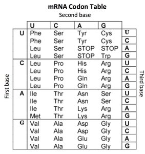
Key Point: The start codon (AUG) signals the beginning of translation and codes for methionine.
Key Point: Stop codons (UAA, UAG, UGA) signal the end of translation.
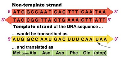
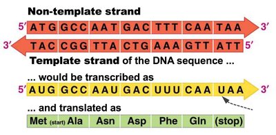
Example: The mRNA sequence 5’ AUG GCA AAU GAC UUU CAA UAA 3’ is translated as Met-Ala-Asn-Asp-Phe-Gln-(stop).
The Genetic Code and Codons
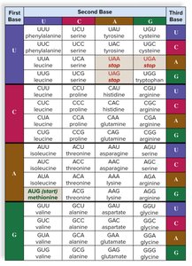
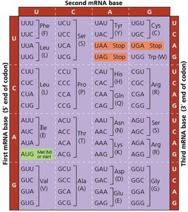
The genetic code consists of 64 codons, each made up of three nucleotides. Codons are read sequentially by the ribosome, and each codon corresponds to a specific amino acid or a stop signal. The code is redundant, meaning multiple codons can code for the same amino acid.
Key Point: There are 20 common amino acids and 64 possible codons.
Key Point: Codons are read in the 5’ → 3’ direction on the mRNA.
Key Point: The start codon is AUG (methionine).
Key Point: Stop codons are UAA, UAG, and UGA.
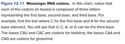
Example: Codons CAU and CAC both code for histidine; CAA and CAG code for glutamine.
Transcription and Translation: From DNA to Protein
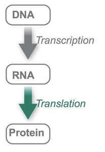
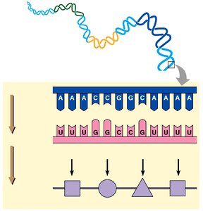
Transcription is the process by which DNA is copied into mRNA. The mRNA then serves as a template for translation. The coding strand of DNA matches the mRNA sequence (except T is replaced by U), while the template strand is used to synthesize the mRNA.
Key Point: The non-template (coding) strand of DNA is similar to the mRNA sequence.
Key Point: The template strand is used to synthesize mRNA.
Key Point: mRNA is translated into a sequence of amino acids.
Example: DNA sequence 5’ ATG GCA AAT GAC TTT CAA TAA 3’ is transcribed to mRNA 5’ AUG GCA AAU GAC UUU CAA UAA 3’ and translated to Met-Ala-Asn-Asp-Phe-Gln-(stop).
Components of Translation
Types of RNA
Three main types of RNA are involved in translation: messenger RNA (mRNA), transfer RNA (tRNA), and ribosomal RNA (rRNA). Each plays a distinct role in the synthesis of proteins.
mRNA: Carries the genetic code from DNA to the ribosome.
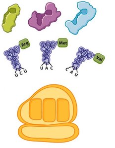
tRNA: Brings amino acids to the ribosome and matches them to the codons in mRNA via its anticodon.
rRNA: Forms the core of the ribosome and catalyzes peptide bond formation.
tRNA Structure and Function
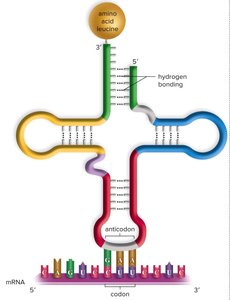
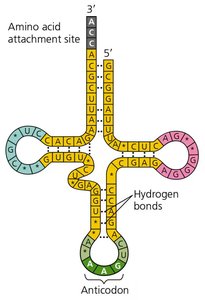
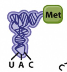
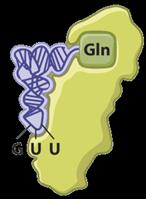
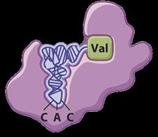
Transfer RNA (tRNA) is a small RNA molecule that carries a specific amino acid and recognizes the corresponding codon on the mRNA through its anticodon. The anticodon is antiparallel and complementary to the mRNA codon.
Key Point: Each tRNA is charged with a specific amino acid by an aminoacyl-tRNA synthetase.
Key Point: The anticodon of tRNA base pairs with the codon of mRNA.
Key Point: tRNA structure includes an amino acid attachment site and an anticodon loop.
Example: tRNA with anticodon UAC carries methionine and pairs with the start codon AUG.
Aminoacyl-tRNA Synthetases
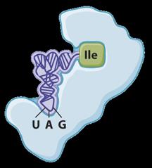
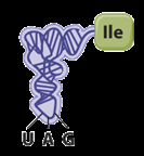
Aminoacyl-tRNA synthetases are enzymes that attach the correct amino acid to its corresponding tRNA. There is a specific synthetase for each amino acid.
Key Point: Each amino acid has a unique synthetase enzyme.
Key Point: The enzyme recognizes both the amino acid and the tRNA.
Example: Isoleucine tRNA synthetase attaches isoleucine to its tRNA.
Ribosome Structure and Function
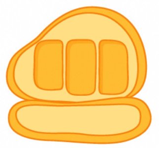
The ribosome is a complex molecular machine composed of rRNA and proteins. It has two subunits (large and small) and three binding sites for tRNA: the A (aminoacyl), P (peptidyl), and E (exit) sites. The ribosome catalyzes peptide bond formation and moves along the mRNA during translation.
Key Point: The ribosome reads mRNA codons and matches them with tRNA anticodons.
Key Point: The ribosome has A, P, and E sites for tRNA binding.
Key Point: Peptide bonds are formed between amino acids in the P and A sites.

Mechanism of Translation
Initiation
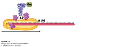
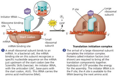
Translation initiation begins when the small ribosomal subunit binds to the mRNA at the 5’ cap and scans for the start codon (AUG). The initiator tRNA carrying methionine binds to the start codon. The large ribosomal subunit then joins, forming the complete initiation complex.
Key Point: Initiation factors help assemble the initiation complex.
Key Point: The start codon is recognized by the initiator tRNA (anticodon UAC).
Key Point: The P site holds the initiator tRNA during initiation.
Elongation
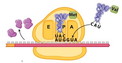
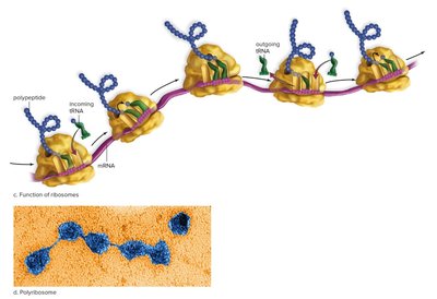
During elongation, the ribosome moves along the mRNA, matching each codon with the appropriate tRNA. Peptide bonds are formed between amino acids, and the polypeptide chain grows. The ribosome shifts one codon at a time, moving tRNAs through the A, P, and E sites.
Key Point: Elongation factors assist in the movement and binding of tRNAs.
Key Point: Peptide bonds are catalyzed by the ribosome (ribozyme activity).
Key Point: The ribosome moves in the 5’ → 3’ direction along the mRNA.
Example: The tRNA in the A site receives the growing polypeptide chain from the tRNA in the P site.
Termination
Termination occurs when a stop codon enters the A site of the ribosome. Release factors bind to the stop codon, causing the polypeptide to be released from the tRNA and the ribosome to dissociate.
Key Point: Stop codons do not code for amino acids.
Key Point: Release factors recognize stop codons and trigger termination.
Key Point: The completed polypeptide is released for folding and further processing.
Protein Targeting and Synthesis Locations
Protein Synthesis in the Cytoplasm and Rough ER
Proteins are synthesized in two main locations: free ribosomes in the cytoplasm and ribosomes bound to the rough endoplasmic reticulum (RER). Proteins destined for secretion or membrane insertion are synthesized on the RER.
Key Point: Free ribosomes synthesize proteins for the cytoplasm.
Key Point: Ribosomes bound to the RER synthesize proteins for secretion, membranes, or organelles.
Key Point: Signal sequences direct ribosomes to the RER.
Transmembrane Protein Synthesis
Transmembrane proteins are synthesized on the RER and inserted into the lipid bilayer. The transmembrane domain, typically composed of hydrophobic amino acids, anchors the protein in the membrane.
Key Point: Transmembrane domains embed proteins in the lipid bilayer.
Key Point: Proteins are processed through the secretory pathway: ER → Golgi → destination.
Summary Table: Codons and Amino Acids
Codon | Amino Acid |
|---|---|
AUG | Methionine (Start) |
UAA, UAG, UGA | Stop |
CAU, CAC | Histidine |
CAA, CAG | Glutamine |
GCA | Alanine |
UUU | Phenylalanine |
GAC | Aspartic acid |
Key Equations and Concepts
Number of possible codons:
Direction of translation: mRNA is read 5’ → 3’
Polypeptide synthesis: N-terminus → C-terminus
Practice Questions
Each of the 20 common amino acids is specified by: C. one or more codons
The tRNA anticodon: D. all of the above (antiparallel, hydrogen bonds, base pairs with codon)
The mRNA is: D. A and B are true (read and made in 5’ to 3’ direction)
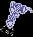
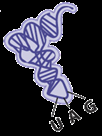
If translation begins with the first start codon in the mRNA below, the second codon would be: A. 5’-CAC-3’
Which amino acid will be attached to this tRNA (anticodon 3’ CGU 5’)? Alanine
Summary
Translation decodes mRNA's triplet code (codons) to amino acids, converting nucleic acid information into protein structure and function. This process is central to gene expression and cellular function.
