 Indietro
Study Guide: The Nervous System (Medical Terminology)
Guida di studio - Note intelligenti
 Appunti personalizzati basati sui tuoi materiali, ampliati con definizioni chiave, esempi e contesto.
The Nervous System
Overview and Functional Roles
The nervous system is a complex network responsible for sensing environmental changes, integrating information, and producing motor responses. It consists of the brain, spinal cord, and nerves, and is divided into two main parts: the central nervous system (CNS) and peripheral nervous system (PNS).
Sensory Input: Sensory neurons detect stimuli and transmit information to the CNS (afferent pathway).
Integration: Interneurons process information and determine appropriate responses.
Motor Output: Motor neurons send signals to effectors (muscles or glands) to produce movement (efferent pathway).
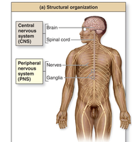
Central vs. Peripheral Nervous System
The nervous system is structurally and functionally divided as follows:
Central Nervous System (CNS): Composed of the brain and spinal cord. Responsible for processing and integrating information.
Peripheral Nervous System (PNS): Composed of nerves and ganglia. Transmits information between the CNS and the rest of the body.
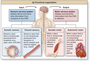
Sensory vs. Motor Nervous System
The nervous system is functionally organized into sensory and motor divisions:
Sensory Nervous System: Detects and transmits information from receptors to the CNS.
Motor Nervous System: Transmits information from the CNS to effectors.
Somatic Sensory: Consciously perceived input (e.g., eyes, skin).
Visceral Sensory: Not consciously perceived input (e.g., internal organs).
Somatic Motor: Voluntary motor output to skeletal muscle.
Autonomic Motor: Involuntary motor output to cardiac muscle, smooth muscle, and glands.
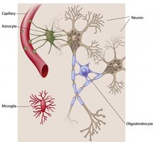
Cell Types of the Nervous System
Neurons and Glial Cells
The nervous system contains two major cell types:
Neurons: Primary cells that transmit information via electrical signals.
Glial Cells (Neuroglia): Support and protect neurons. Types include astrocytes, oligodendrocytes, Schwann cells, and microglia.
Structures of a Neuron
Neurons have specialized structures for communication:
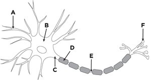
Cell Body (Soma): Contains organelles and nucleus.
Dendrites: Receive incoming signals.
Axon: Conducts action potentials away from the cell body.
Synaptic Terminals: Release neurotransmitters to target cells.
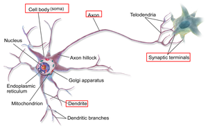
Practice: Identify Neuron Structures
A: Dendrite
B: Cell body
C: Axon hillock
D: Myelin sheath
E: Axon
F: Synaptic terminals
Glial Cells and Myelination
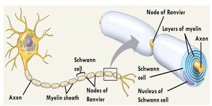
Schwann Cells: Form myelin sheath around axons in the PNS, increasing action potential speed.
Myelinated Neurons: Have myelin sheath; constitute white matter.
Unmyelinated Neurons: Lack myelin sheath; constitute gray matter.
Clinical View: Multiple Sclerosis (M.S.)
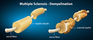
Definition: Autoimmune disorder where oligodendrocytes are attacked, damaging the myelin sheath.
Consequences: Regeneration possible, but scarring of axons can cause permanent deficits.
Nerves and Their Structure
Cranial vs. Spinal Nerves
Cranial Nerves: 12 pairs, extend from the brain.
Spinal Nerves: 31 pairs, extend from the spinal cord.
Structure of Nerves
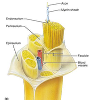
Nerves are bundles of axons wrapped in connective tissue:
Endoneurium: Surrounds individual axons.
Perineurium: Surrounds fascicles (bundles of axons).
Epineurium: Surrounds the entire nerve.
Spinal Reflexes
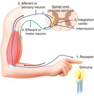
Reflex Arc
Spinal reflexes are rapid, involuntary responses initiated by the spinal cord.
Stimulus activates receptor.
Sensory (afferent) neuron transmits signal.
Integration center (interneuron) processes information.
Motor (efferent) neuron transmits response.
Effector (muscle or gland) acts.
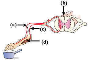
Practice: Reflex Arc Structure Identification
(a): Afferent neuron
(b): Integration center
(c): Efferent neuron
(d): Effector (muscle)
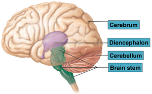
Major Brain Regions
Four Main Brain Regions
The brain is divided into four major regions, each with distinct functions and substructures:
Cerebrum
Diencephalon
Cerebellum
Brain Stem
Cerebrum
Largest and most superior part of the brain.
Divided into left and right hemispheres by the longitudinal fissure.
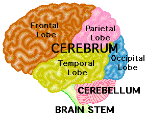
Each hemisphere has five lobes: frontal, parietal, occipital, temporal, and insula.
Gray matter: Superficial, processes information (cerebral cortex, cerebral nuclei).
White matter: Deep, transmits information (tracts such as corpus callosum).
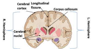
Surface Features of Cerebrum
Sulci: Depressions
Gyri: Ridges
Fissures: Deep depressions
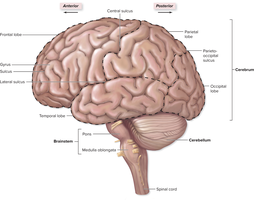
Functional Areas of Cerebral Cortex
Frontal Lobe: Somatic motor control, learning, decision making, personality.
Parietal Lobe: Somatic sensory processing, spatial awareness.
Occipital Lobe: Vision processing.
Temporal Lobe: Hearing, memory (hippocampus).
Decussation and Lateralization
Decussation: Axons cross over; left hemisphere controls right side of body and vice versa.
Lateralization: Hemispheres specialize in certain tasks (e.g., left hemisphere for complex reasoning in most people).
Diencephalon
Epithalamus and Pineal Gland: Secrete melatonin, regulate circadian rhythm.
Thalamus: Relay and filter information from brainstem to cerebrum.
Hypothalamus: Master regulator of homeostasis, controls autonomic and endocrine systems, regulates temperature, hunger, thirst.
Brainstem
Midbrain: Visual and auditory reflexes.
Pons: Breathing, urination, cranial nerve functions.
Medulla Oblongata: Autonomic control centers for heart rate, blood vessel diameter, and breathing.
Cerebellum
Second largest brain region.
Fine-tunes movement commands, stores learned movement memories.
Brain Protective Structures
Meninges
Pia Mater: Innermost layer, directly on brain surface.
Arachnoid Mater: Middle layer, above subarachnoid space (contains CSF).
Dura Mater: Outermost layer, tough connective tissue.
Brain Ventricles and Cerebrospinal Fluid (CSF)
Four Ventricles: Lateral, third, fourth; produce and contain CSF.
Choroid Plexus: Produces CSF.
CSF: Cushions, nourishes, removes wastes, and provides immune support.
Old CSF is reabsorbed by arachnoid membranes.
Disorders Related to Meninges/Ventricles/CSF
Meningitis: Inflammation of meninges due to infection.
Epi/Subdural Hematoma: Blood pooling from ruptured vessels in meningeal layers.
Hydrocephalus: Excess CSF accumulation, causes swelling.
Blood-Brain Barrier
Highly selective barrier: Regulates entry of substances from blood to brain tissue.
Features thicker vessel walls, tight junctions, and astrocyte foot processes.
Autonomic Nervous System (ANS)
Sympathetic vs. Parasympathetic Nervous System
The ANS provides unconscious motor control of internal organs, smooth muscle, and glands.
Sympathetic Nervous System: Activates during stress (fight or flight), increases alertness and physical activity. Main neurotransmitter: epinephrine.
Parasympathetic Nervous System: Active during rest (rest and digest), promotes relaxation and digestion. Main neurotransmitter: acetylcholine.
Major Effects of Sympathetic and Parasympathetic Nervous System
Sympathetic: Dilates pupils, bronchodilation, increases heart rate, stimulates adrenal gland, redirects blood flow to muscles.
Parasympathetic: Constricts pupils, promotes digestion, slows heart rate, stimulates glandular secretions.
Summary Table: CNS vs. PNS
Feature | CNS | PNS |
|---|---|---|
Components | Brain, Spinal Cord | Nerves, Ganglia |
Function | Integration, Processing | Transmission, Communication |
Cell Types | Neurons, Glial Cells | Neurons, Schwann Cells |
Key Terms
Neuron: Cell that transmits electrical signals.
Glial Cell: Supportive cell in nervous system.
Myelin Sheath: Insulating layer around axons.
Reflex Arc: Pathway for rapid, involuntary response.
Meninges: Protective layers around brain and spinal cord.
Blood-Brain Barrier: Selective barrier protecting brain tissue.
Sympathetic/Parasympathetic: Branches of ANS controlling stress and rest responses.
Practice Questions
What type of neuron activates sweat glands? Autonomic motor neuron
What type of CNS injury causes loss of vision? Occipital lobe
What type of CNS injury causes loss of concentration/personality? Frontal lobe
Which brain region coordinates and fine-tunes movements? Cerebellum
Which is not a component of the blood-brain barrier? Microglia
Which is not an effect of the sympathetic nervous system? Storage of fuel molecules in the liver
Equations and Formulas
Resting Membrane Potential:
Action Potential Propagation Speed:
Additional info: Equations provided for academic completeness; not directly in original notes.
