 Indietro
Urinary System: Medical Terminology and Anatomy Study Guide
Guida di studio - Note intelligenti
 Appunti personalizzati basati sui tuoi materiali, ampliati con definizioni chiave, esempi e contesto.
Urinary System Overview
Introduction
The urinary system, also known as the excretory, genitourinary (GU), or urogenital (UG) system, is responsible for producing, storing, and eliminating urine. It plays a vital role in maintaining homeostasis by removing waste products and regulating fluid and electrolyte balance.
Organs: Two kidneys, two ureters, one bladder, one urethra
Primary Functions: Extraction of wastes, urine formation, urine transport, and elimination
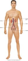
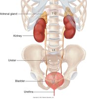
Anatomy and Physiology of the Urinary System
Kidneys
The kidneys are purplish-brown, bean-shaped organs located in a retroperitoneal position from T12 to L3 vertebrae. They are essential for filtering blood, removing wastes, and maintaining fluid and electrolyte balance.
Functions:
Remove wastes and extra fluid
Remove acid produced by cells
Maintain water, salts, and minerals balance
Produce urine
Produce hormones: erythropoietin, renin, angiotensin
Convert vitamin D to calcitriol
External Structure: Concave and convex borders; hilum (notch); renal pelvis (collecting area)
Internal Structure:
Cortex: Outer layer with arteries, veins, convoluted tubules, glomerular capsules
Medulla: Inner portion with renal pyramids and papillae projecting into calyces
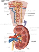
Nephron Structure and Function
Nephrons are the structural and functional units of the kidney, responsible for regulating, controlling, and removing waste products from blood plasma.
Renal Corpuscle: Glomerulus (capillary cluster) surrounded by Bowman capsule
Renal Tubule: Proximal tubule, nephron loop (loop of Henle), distal tubule, collecting tubule
Vital Functions:
Regulate fluid balance
Reabsorb water and electrolytes
Remove metabolic wastes (urea, uric acid, creatinine, ketone bodies)
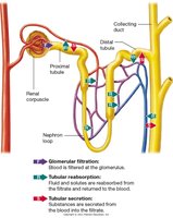
Glomerular Filtration: Filters excess fluid and waste into urine-collecting tubules
Tubular Reabsorption: Allows selected electrolytes and water back into blood
Tubular Secretion: Removes excess acid and other substances
Equation: Glomerular Filtration Rate (GFR) is a key measure of kidney function:
Ureters
Each kidney has a ureter, a narrow muscular tube that drains urine to the bladder. The ureter wall consists of three layers: mucous membrane, smooth muscle, and fibrous tissue.
Urinary Bladder
The bladder is a muscular, membranous sac in the anterior pelvic cavity, serving as a reservoir for urine. It consists of four layers: epithelium, smooth muscle, longitudinal muscle (detrusor urinae), and fibrous layer. The trigone is a triangular area near the base.
Urethra
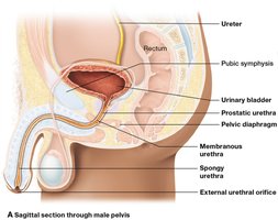
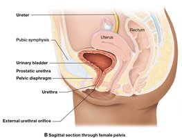
The urethra is a musculomembranous tube extending from the bladder to the outside. The external opening is the urinary meatus.
Male Urethra: ~20 cm, conveys urine and semen, divided into prostatic, membranous, and penile sections
Female Urethra: ~4 cm, conveys urine only, meatus between clitoris and vaginal opening
Urine Formation and Properties
Urine Production
Urine is produced in three main steps: filtration, reabsorption, and secretion. The nephron is the site of these processes.
Urine is 95% water and 5% solid substances
Normal adult voids 1000–1500 mL daily; urge to void at 300–350 mL
Normal urine: clear, yellow to amber, faint odor, specific gravity 1.003–1.030, slightly acidic pH
Urinalysis
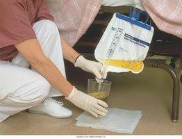
Urinalysis (UA) is a laboratory test evaluating physical, chemical, and microscopic properties of urine. It is a valuable diagnostic tool for detecting metabolic, kidney, and liver disorders, as well as urinary tract infections (UTIs).
Freshly voided specimens provide accurate results
Bacteriologic cultures require catheterization
Common Disorders and Diagnostic Procedures
Urinary Tract Infections (UTIs)
UTIs are common, especially in children and sexually active women. Most are caused by Escherichia coli, Klebsiella, and Proteus.
Infants: Fever, weight loss, nausea, increased urination, strong-smelling urine, diaper rash, failure to thrive
Older Children: Frequent/painful urination, abdominal pain, hematuria, fever, chills, bedwetting
Cystitis and Interstitial Cystitis
Cystitis is common, especially in women due to shorter urethra. Interstitial cystitis is a painful inflammation of the bladder wall, mostly affecting women, and does not respond well to antibiotics.
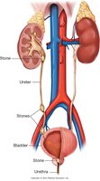
Urinary Calculi (Stones)
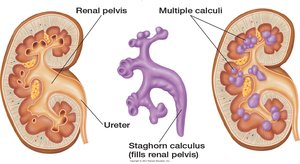
Renal calculi (kidney stones) can form in the renal pelvis, ureter, bladder, or urethra, causing obstruction and pain.
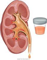
Hematuria
Hematuria is the presence of blood in urine, often indicating infection, stones, or trauma.
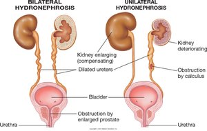
Hydronephrosis
Hydronephrosis is the swelling of a kidney due to a build-up of urine, often caused by obstruction.
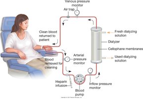
Dialysis
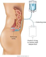
Dialysis is a treatment for kidney failure, using machines or peritoneal cavity to filter blood.
Hemodialysis: Blood is filtered through a machine
Peritoneal Dialysis: Uses the peritoneal cavity as a filter
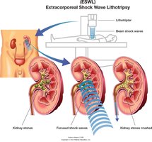
Extracorporeal Shock Wave Lithotripsy (ESWL)
ESWL is a non-invasive procedure using shock waves to break kidney stones into smaller fragments for easier passage.
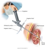
Percutaneous Ultrasonic Lithotripsy
A nephroscope is inserted into the renal pelvis, and ultrasound waves fragment stones, which are then removed.
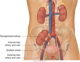
Kidney Transplantation
Transplantation involves placing a healthy kidney from a donor into a patient with kidney failure.
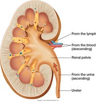
Routes of Infection for Pyelonephritis
Pyelonephritis is a kidney infection that can occur via ascending infection from the urinary tract or through the bloodstream.
Medical Terminology
Key Terms and Word Building
Albuminuria: Presence of albumin (protein) in urine
Cystectomy: Surgical excision of the bladder
Dysuria: Difficult or painful urination
Nephrolithiasis: Condition of kidney stones
Oliguria: Scanty urine production
Urologist: Specialist in urinary system
Drug Highlights
Common Drug Classes
Diuretics: Promote urine production
Urinary tract antibacterials: Treat infections
Urinary tract antiseptics: Prevent infection
Diagnostic and Laboratory Tests
Common Tests
Blood Urea Nitrogen (BUN): Measures urea in blood
Creatinine Clearance: Assesses glomerular filtration rate
Urine Culture: Detects microorganisms
Cystoscopy: Visual examination of bladder and urethra
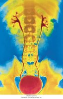
Intravenous Pyelography (IVP): X-ray visualization of urinary tract
Renal Biopsy: Tissue removal for analysis
Retrograde Pyelography: X-ray with contrast medium injected backward
Serum Creatinine: Measures creatinine in blood
Ultrasonography: Uses sound waves to visualize kidneys
Additional Information
Urine formation begins in fetal development at 10 weeks gestation
Newborn kidneys are immature and cannot concentrate urine
Glomerular filtration and reabsorption mature by age 1–2 years
Older adults may experience reduced bladder capacity and urge incontinence
Benign prostatic hyperplasia (BPH) and prostate cancer can cause incontinence in men
Summary Table: Normal and Abnormal Constituents of Urine
Purpose: Classification of urine components for diagnostic purposes.
Constituent | Normal | Abnormal |
|---|---|---|
Water | Present | Absent (dehydration) |
Urea | Present | Excess (renal dysfunction) |
Protein | Absent | Present (proteinuria) |
Glucose | Absent | Present (glycosuria) |
Bilirubin | Absent | Present (biliuria) |
Blood | Absent | Present (hematuria) |
Nitrites | Absent | Present (nitrituria) |
Ketones | Absent | Present (ketonuria) |
Leukocytes | Absent | Present (infection) |
Crystals | Absent | Present (calculi) |
Additional info: Table entries inferred from standard urinalysis parameters.
Abbreviations and Acronyms
Common abbreviations used in urinary system terminology include:
UA: Urinalysis
BUN: Blood Urea Nitrogen
IVP: Intravenous Pyelogram
GFR: Glomerular Filtration Rate
UTI: Urinary Tract Infection
