 Indietro
Cell Structure and Function in Microbiology: Prokaryotic and Eukaryotic Cells
Guida di studio - Note intelligenti
 Appunti personalizzati basati sui tuoi materiali, ampliati con definizioni chiave, esempi e contesto.
Processes of Life
Characteristics of Living Cells
All living organisms share several fundamental processes that define life. These include:
Growth: An increase in size or number of cells.
Reproduction: The ability to produce new cells or organisms, either sexually or asexually.
Responsiveness: The capacity to respond to environmental stimuli.
Metabolism: The sum of all chemical reactions within the organism, including energy production and biosynthesis.
Cell Types: Prokaryotes vs. Eukaryotes
Overview of Cell Types
Cells are classified into two main types based on their structural characteristics: prokaryotic and eukaryotic cells.
Prokaryotes:
Lack a nucleus and membrane-bound organelles.
Generally smaller (~1.0 µm in diameter) and simpler in structure.
Include Bacteria and Archaea.
Eukaryotes:
Possess a true nucleus and various internal membrane-bound organelles.
Larger (10–100 µm in diameter) and more complex.
Include Algae, Protozoa, Fungi, Animals, and Plants.
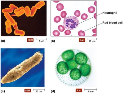
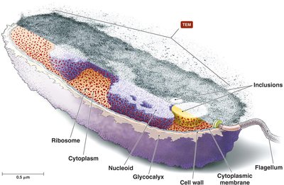
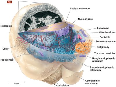
Relative Sizes of Cells
Cells and microorganisms vary greatly in size, from viruses (0.3 µm) to chicken eggs (47,000 µm). Understanding these size differences is crucial for microscopy and laboratory techniques.
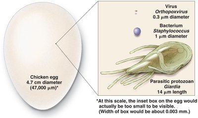
External Structures of Bacterial Cells
Glycocalyces
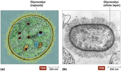
The glycocalyx is a gelatinous, sticky substance surrounding the outside of some bacterial cells. It is composed of polysaccharides, polypeptides, or both, and serves as a protective layer.
Capsule: Organized, firmly attached to the cell surface, may prevent recognition by host immune systems.
Slime Layer: Loosely attached, water-soluble, aids in adherence to surfaces.
Flagella
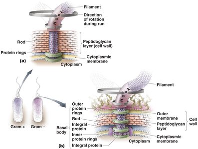
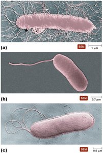
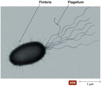
Flagella are long, whip-like structures responsible for bacterial motility. Not all bacteria possess flagella.
Structure: Composed of a filament, hook, and basal body. The basal body anchors the flagellum to the cell wall and membrane.
Function: Rotation of the flagellum propels the bacterium. Movement can be in response to stimuli (taxis), with alternating runs and tumbles.
Fimbriae and Pili
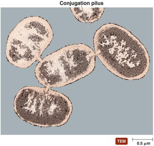
Fimbriae are short, bristlelike projections that help bacteria adhere to surfaces and each other, playing a key role in biofilm formation. Pili (or conjugation pili) are longer, less numerous, and mediate the transfer of DNA between cells (conjugation).
Bacterial Cell Walls
Structure and Function
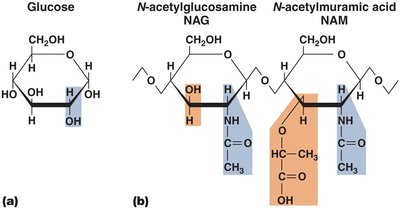
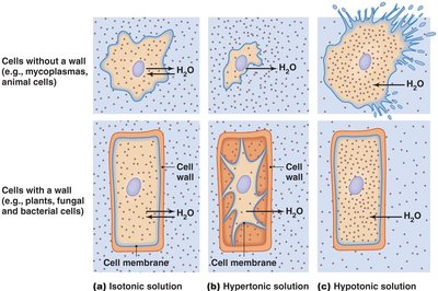
The bacterial cell wall provides structural support, shape, and protection from osmotic pressure. It is primarily composed of peptidoglycan, a polymer of sugars and amino acids.
Cell wall composition is a major target for antibiotics.
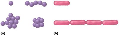
Cell wall structure determines the characteristic shape of bacteria (cocci, bacilli, etc.).
Gram-Positive vs. Gram-Negative Cell Walls
Gram-Positive:
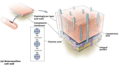
Thick peptidoglycan layer
Contains teichoic acids
Stains purple in Gram stain
Some have mycolic acid (acid-fast bacteria)
Gram-Negative:
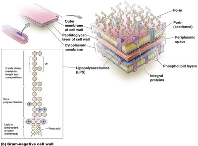
Thin peptidoglycan layer
Outer membrane with lipopolysaccharide (LPS)
Stains pink in Gram stain
Outer membrane can impede antibiotic treatment
Bacterial Cytoplasmic Membrane
Structure
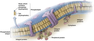
The cytoplasmic membrane is a phospholipid bilayer with embedded proteins, described by the fluid mosaic model. It acts as a selective barrier and is involved in energy generation and transport.
Functions
Energy storage and harvesting (in photosynthetic bacteria).
Selective permeability: Only certain substances can cross freely; others require transport proteins.
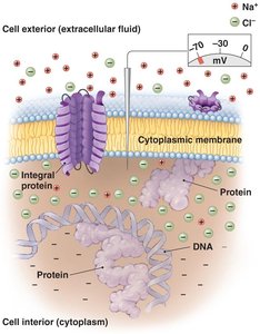
Maintains concentration and electrical gradients.
Transport Mechanisms
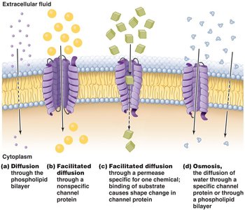
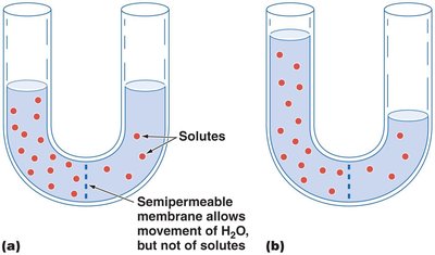
Passive Processes (no energy required):
Diffusion
Facilitated diffusion
Osmosis
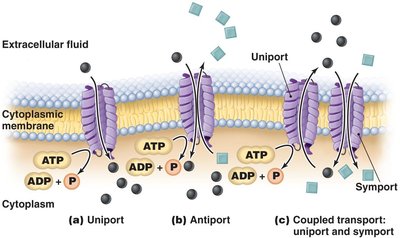
Active Processes (energy required):
Active transport (uniport, antiport, symport)
Group translocation (substance is chemically modified during transport)
Cytoplasm of Bacteria
Components
Cytosol: Liquid portion containing dissolved substances.
Inclusions: Reserve deposits of chemicals.
Endospores: Dormant, resistant structures formed by some bacteria for survival in harsh conditions.
Ribosomes: Sites of protein synthesis (70S in prokaryotes).
Cytoskeleton: Maintains cell shape.
Archaeal Cell Structure
External Structures
Glycocalyces: Involved in biofilm formation and adherence.
Flagella: Structurally different from bacterial flagella.
Fimbriae and Hami: Attachment structures; hami are unique to archaea.
Cell Walls and Membranes
Most archaea have cell walls (no peptidoglycan; contain specialized polysaccharides and proteins).
All have cytoplasmic membranes that maintain gradients and control transport.
Cytoplasm
Similar to bacteria (70S ribosomes, circular DNA, cytoskeleton).
Differences: Unique ribosomal proteins, metabolic enzymes, and genetic code more similar to eukaryotes.
Eukaryotic Cell Structure
External Structures
Glycocalyces: Less organized than prokaryotic capsules; function in cell recognition, adhesion, and protection.
Cell Walls and Membranes
Present in fungi, algae, plants, and some protozoa; composed of cellulose, chitin, glucomannan, or other polysaccharides.
All eukaryotes have a cytoplasmic membrane with steroid lipids and membrane rafts.
Motility Structures
Flagella: Structurally distinct from prokaryotic flagella; composed of microtubules, undulate rather than rotate.
Cilia: Shorter, more numerous; move cells or substances across surfaces.
Nonmembranous Organelles
Ribosomes: Larger (80S) than prokaryotic ribosomes; composed of 60S and 40S subunits.
Cytoskeleton: Network of microtubules, microfilaments, and intermediate filaments; maintains cell shape and anchors organelles.
Centrioles and Centrosome: Involved in cell division and formation of cilia/flagella.
Membranous Organelles
Nucleus: Contains DNA, nucleolus (site of RNA synthesis), surrounded by nuclear envelope with pores.
Endoplasmic Reticulum (ER): Network of membranes; rough ER (with ribosomes) synthesizes proteins, smooth ER synthesizes lipids.
Golgi Body: Modifies, sorts, and packages proteins and lipids for secretion or delivery to other organelles.
Lysosomes, Peroxisomes, Vacuoles, Vesicles: Involved in storage, digestion, and detoxification.
Mitochondria: Site of ATP production; contain their own DNA and 70S ribosomes.
Chloroplasts: Site of photosynthesis in plants and algae; contain DNA and 70S ribosomes.
Endosymbiotic Theory
The endosymbiotic theory proposes that eukaryotic cells originated through a symbiotic relationship between early prokaryotic cells. Aerobic prokaryotes became mitochondria, and photosynthetic prokaryotes became chloroplasts. This theory is supported by the presence of 70S ribosomes and circular DNA in these organelles, but it is not universally accepted.
