 Indietro
Innate Immunity: Principles and Clinical Applications
Guida di studio - Note intelligenti
 Appunti personalizzati basati sui tuoi materiali, ampliati con definizioni chiave, esempi e contesto.
Innate Immunity: Principles and Clinical Applications
Overview of the Immune System
The immune system is a complex network that protects the body from pathogens through coordinated physiological processes. Immunity is divided into two main branches: innate immunity and adaptive immunity. Both branches share the ability to recognize diverse pathogens, eliminate invaders, and distinguish between self and foreign antigens.
Innate Immunity: Inborn, nonspecific defense mechanisms present in all eukaryotes. Provides immediate, generalized protection.
Adaptive Immunity: Found only in vertebrates, matures over time, and tailors responses to specific pathogens. Exhibits memory and typically takes 4–7 days to fully activate.
Immunity refers to specific protection conferred by adaptive responses, while susceptibility indicates vulnerability to infection.
Collaboration Between Innate and Adaptive Immunity
Immunity operates through three lines of defense: barrier defenses, cellular and molecular defenses, and adaptive defenses. These lines collaborate to provide comprehensive protection.
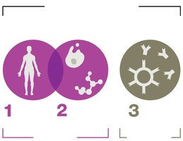
Role of Normal Microbiota in Immune Responses
Normal microbiota are symbiotic microbes that inhabit the body, shaping immune responses and excluding pathogens. They help train the immune system to tolerate nonpathogens and self-tissues. Disruptions in microbiota can lead to immune confusion, potentially contributing to allergies and autoimmunity (the hygiene hypothesis).
Germ-free animal studies show underdeveloped immune systems, highlighting the importance of microbiota in immune development.
First-Line Defenses
General Function and Categories
First-line defenses aim to prevent pathogen entry and are categorized as mechanical, chemical, and physical barriers.
Mechanical Barriers: Flushing, rinsing, and trapping actions (e.g., tears, urine, saliva, mucus membranes, mucociliary escalator).
Chemical Barriers: Molecules that attack microbes or create hostile environments (e.g., lysozyme, stomach acid, fatty acids, antimicrobial peptides).
Physical Barriers: Structural blockades such as epithelial tissue and skin.
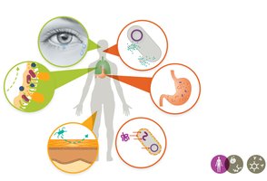
Mechanical Barriers
Tears wash debris from eyes.
Urine flushes microbes from the urinary tract.
Saliva limits microbial adherence.
Mucus membranes trap microbes at body entrances and internal linings.
Mucociliary escalator moves trapped particles out of the lungs.
Chemical Barriers
Lysozyme: Enzyme found in tears, saliva, mucus, and breast milk; breaks down bacterial cell walls.
Antimicrobial Peptides (AMPs): Proteins that destroy a wide range of pathogens by disrupting membranes or targeting intracellular components.
Defensins: A class of AMPs that rapidly kill invaders by inserting into cell membranes.
Other examples: Hydrochloric acid in the stomach, fatty acids in sweat and earwax, dry and acidic skin environment.
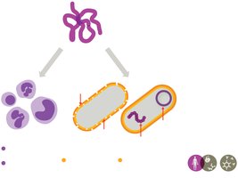
Physical Barriers
Epithelial tissue: Lines all body cavities and entrances, forming a main physical barrier.
Skin: Epidermis consists of tightly packed dead cells enriched with proteins and lipids, creating a water-resistant layer.
Second-Line Defenses and the Lymphatic System
Introduction to Second-Line Defenses
When first-line defenses are breached, second-line defenses—comprising molecular factors and leukocytes—are activated to eliminate pathogens.
The Lymphatic System
The lymphatic system collects, circulates, and filters body fluids, playing a crucial role in immune surveillance and response.
Lymph: Interstitial fluid taken up by lymphatic capillaries, filtered in lymph nodes, and returned to the bloodstream.
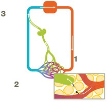
Primary and Secondary Lymphoid Tissues
Primary lymphoid tissues: Sites of leukocyte production and maturation (thymus for T cells, bone marrow for B cells and other blood cells).
Secondary lymphoid tissues: Filter lymph and sample antigens (lymph nodes, spleen, mucosa-associated lymphoid tissue—MALT).
Leukocytes (White Blood Cells)
Leukocytes are essential for all immune responses and are classified as granulocytes (with cytoplasmic granules) or agranulocytes (without granules).
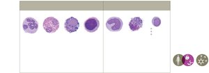
Granulocytes: Neutrophils, eosinophils, basophils, mast cells
Agranulocytes: Monocytes (macrophages), dendritic cells, lymphocytes (NK cells, B cells, T cells)
White Blood Cell Counts in Clinical Diagnosis
Differential WBC count: Measures proportions of different leukocytes to diagnose infections or immune disorders.
Leukocytosis: Elevated leukocyte count, often indicating infection.
Cellular Second-Line Defenses
Granulocytes
Neutrophils: Most abundant, first responders, phagocytize pathogens, release AMPs. Elevated in acute bacterial infections.
Eosinophils: Combat parasites and allergens, moderate phagocytic activity, release enzymes and toxins.
Basophils: Release histamine, involved in allergic responses and fighting parasites.
Mast cells: Reside in tissues, release histamine, conduct phagocytosis, common near body openings.
Agranulocytes
Monocytes: Largest WBCs, mature into macrophages in tissues, increase in chronic infections and inflammation.
Macrophages: Highly phagocytic, can be fixed or wandering, activate adaptive immunity.
Dendritic cells: Highly phagocytic, patrol tissues, modulate immune responses.
Lymphocytes
Natural Killer (NK) cells: Innate immunity against viruses, bacteria, parasites, and tumor cells.
B cells and T cells: Coordinate adaptive immune responses (covered in detail in Chapter 12).
Molecular Second-Line Defenses
Key Roles and Molecules
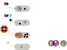
Leukocytes release molecules that recruit other immune cells, restrict pathogen growth, trigger fever, and stimulate inflammation.
Cytokines
Cytokines: Signaling proteins for cell communication and immune coordination. Overproduction can cause a cytokine storm.
Chemokines: Attract WBCs to infection sites (chemotaxis), aid in wound healing and immune activation.
Interleukins (ILs): Regulate inflammation, stimulate immune responses, hematopoiesis, and apoptosis.
Interferons (IFNs): Signal viral or tumor presence, induce antiviral defenses, trigger apoptosis, and activate leukocytes.
Tumor Necrosis Factors (TNFs): Stimulate inflammation, kill tumor cells, and induce fever.
Iron-Binding Proteins
Limit free iron to restrict microbial growth (hemoglobin, ferritin, lactoferrin, transferrin).
Pathogens may produce siderophores to steal iron or use alternative metals like manganese.
Complement System
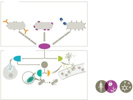
The complement system consists of over 30 proteins that enhance immune responses through a cascade mechanism. Activation occurs via three pathways:
Classical pathway: Triggered by antibodies bound to pathogens.
Alternative pathway: Direct interaction with pathogens, antibody-independent.
Lectin pathway: Initiated by mannose-binding lectin binding to microbial sugars.
All pathways result in:
Opsonization: Tagging invaders for phagocytosis.
Membrane Attack Complex (MAC): Causes cytolysis of pathogens.
Inflammation: Recruitment of immune cells and promotion of inflammatory responses.
Regulation of Complement
Complement proteins are unstable and regulated by proteins (RCAs) to prevent damage to host tissues.
Inflammation and Fever
Inflammation
Inflammation is a key innate response to tissue injury or infection, essential for healing but potentially damaging if unregulated.
Goals: Recruit immune defenses, limit pathogen spread, deliver factors for tissue recovery.
Cardinal signs: Redness, pain, localized heat, swelling, loss of function.
Phases of Inflammation
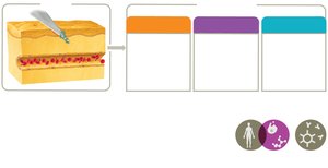
Vascular changes: Vasodilation and increased permeability allow plasma and proteins to enter tissues, forming exudate.
Leukocyte recruitment: Chemoattractants guide leukocytes (neutrophils, then monocytes/macrophages) to the site. Margination and diapedesis enable leukocyte exit from vessels.
Resolution: Chemical signals reduce inflammation, leukocytes undergo apoptosis, exudate is cleared, and tissue repair begins.
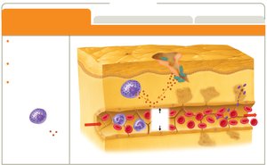
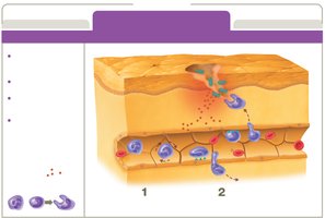
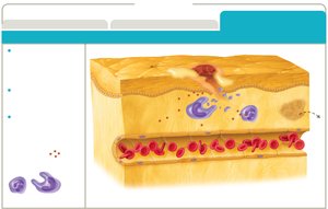
Key Chemical Mediators
Histamine: Released by mast cells, basophils, eosinophils, and platelets; increases vessel permeability.
Kinins: Induce vascular changes, stimulate pain, and amplify inflammation.
Eicosanoids: Promote vascular changes, pain, and fever (e.g., prostaglandins, leukotrienes, thromboxanes).
NSAIDs and SAIDs: Reduce eicosanoid production, limiting inflammation and pain.
Chronic Inflammation
Chronic inflammation results from prolonged immune responses, causing tissue damage and contributing to diseases such as atherosclerosis, cancer, and neurodegenerative disorders.
Fever (Pyrexia)
Fever is a systemic innate response characterized by elevated body temperature, induced by pyrogens (e.g., bacterial toxins, cytokines). The hypothalamus raises the body's temperature set point.
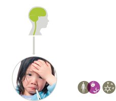
Benefits: Enhances interferon effects, increases phagocyte efficiency, limits pathogen growth, promotes tissue repair.
Low-grade fever is generally protective; high fever can be life-threatening and requires medical intervention.
Antipyretics (e.g., aspirin, ibuprofen, acetaminophen) reduce fever by limiting prostaglandin production in the hypothalamus.
Clinical Case Application
Case Summary: Bacterial Meningitis in a Child
5-year-old with ear infection, fever, and later, bacterial meningitis caused by Streptococcus pneumoniae.
Despite vaccination, infection occurred due to non-vaccine strain.
Low mannose-binding lectin (MBL) levels identified, increasing susceptibility to certain infections.
Clinical management included antibiotics, antipyretics, and early vaccination for other pathogens.
This case illustrates the importance of both first- and second-line defenses, the role of complement (MBL pathway), and the clinical significance of immune deficiencies.
Summary Table: Comparison of Innate and Adaptive Immunity
Feature | Innate Immunity | Adaptive Immunity |
|---|---|---|
Specificity | Nonspecific | Highly specific |
Response Time | Immediate (minutes to hours) | Delayed (days) |
Memory | None | Yes |
Major Components | Barriers, phagocytes, complement, cytokines | B cells, T cells, antibodies |
