 Indietro
Bones and Skeletal Tissues: Structure, Function, and Development
Guida di studio - Note intelligenti
 Appunti personalizzati basati sui tuoi materiali, ampliati con definizioni chiave, esempi e contesto.
Bones and Skeletal Tissues
Skeletal Cartilage
Skeletal cartilage is a resilient tissue that forms much of the embryonic skeleton and persists in adult joints. It is avascular and lacks nerves, relying on diffusion for nutrient supply.
Composition: Primarily water, surrounded by the perichondrium which resists expansion and contains blood vessels.
Types of Cartilage:
Hyaline cartilage: Most abundant; provides support, flexibility, and resilience. Found in articular, costal, respiratory, and nasal cartilages.
Elastic cartilage: Similar to hyaline but with more elastic fibers; found in external ear and epiglottis.
Fibrocartilage: Highly compressed, great tensile strength, contains collagen fibers; found in menisci, pubic symphysis, and intervertebral discs.
Classification of Bones
The human skeleton consists of 206 named bones, divided into axial and appendicular skeletons.
Axial skeleton: Skull, vertebral column, rib cage.
Appendicular skeleton: Limbs, shoulder, hip.
Bone Types:
Long bones: Longer than wide (e.g., humerus).
Short bones: Cube-shaped (e.g., wrist, ankle).
Flat bones: Thin, flattened, curved (e.g., sternum, skull).
Irregular bones: Complex shapes (e.g., vertebrae, pelvis).
Sesamoid bones: Small, round, in tendons (e.g., patella).
Functions of Bones
Bones serve multiple essential functions in the body.
Support: Framework for the body and organs.
Protection: Shields brain, spinal cord, and vital organs.
Movement: Acts as levers for muscles.
Storage: Stores minerals (calcium, phosphate), growth factors, and fat (yellow marrow).
Blood cell formation: Hematopoiesis in red marrow.
Bone Markings
Bone markings are structural features that serve as sites for muscle, ligament, and tendon attachment, joint surfaces, and passageways for blood vessels and nerves.
Bulges, depressions, holes: Facilitate attachment and passage.
Foramen: Holes for vessels and nerves.
Bone Textures
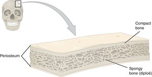
Bones have two main textures: compact and spongy.
Compact bone: Dense outer layer.
Spongy bone: Honeycomb structure of trabeculae.
Gross Anatomy of Bone
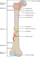
Long bones have distinct anatomical regions and structures.
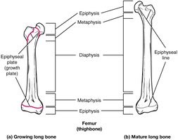
Diaphysis: Tubular shaft, contains medullary cavity (yellow marrow in adults, red in infants).
Epiphysis: Ends of bone, filled with spongy bone and red marrow.
Epiphyseal plate: Growth plate of hyaline cartilage; becomes epiphyseal line after growth.
Compact bone: Forms the wall of diaphysis.
Spongy bone: Fills epiphyses.
Membranes of Bone
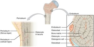
Bones are covered and lined by specialized membranes.
Endosteum: Lines medullary cavity and spongy bone; contains osteoblasts and osteoclasts for growth and repair.
Periosteum: Covers outer surface; contains vessels, nerves, and Sharpey's fibers for attachment.
Articular cartilage: Hyaline cartilage at epiphyses for friction reduction and shock absorption.
Microscopic Structure of Bone: Compact Bone
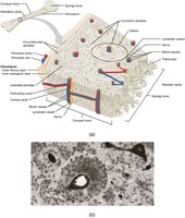
Compact bone is organized into structural units called osteons (Haversian systems).
Osteon: Cylindrical unit with concentric lamellae.
Lamella: Collagen matrix tubes for weight-bearing.
Haversian canal: Central channel for vessels and nerves.
Volkmann's canals: Perforating canals connecting periosteum and Haversian canal.
Osteocytes: Mature bone cells in lacunae.
Canaliculi: Hairlike canals connecting lacunae and central canal.
Microscopic Anatomy of Bone: Spongy Bone
Spongy bone consists of trabeculae aligned along lines of stress, with irregular lamellae and osteocytes.
Trabeculae: Lattice-like structures, no osteons.
Lamellae: Irregularly arranged.
Osteocytes: In lacunae, connected by canaliculi.
Capillaries: In endosteum supply nutrients.
Bone marrow: Fills spaces between trabeculae.
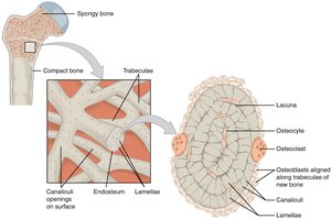
Location of Hematopoietic Tissue (Red Marrow)
Red marrow is the site of blood cell formation.
Infants: Found in medullary cavity and all spongy bone.
Adults: Found in spongy bone of flat bones, head of femur and humerus.
Yellow marrow: Can revert to red marrow in anemia.
Types of Bone Cells
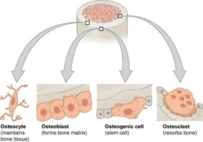
Bone tissue contains four main cell types, each with distinct functions.
Osteocytes: Mature bone cells, maintain bone tissue.
Osteoblasts: Bone-forming cells, synthesize matrix.
Osteoclasts: Large cells, resorb bone matrix.
Osteogenic cells: Stem cells, differentiate into osteoblasts.
Chemical Composition of Bone
Bones are composed of organic and inorganic components.
Hydroxyapatites (mineral salts): 65% of bone mass, mainly calcium phosphates and magnesium, responsible for hardness and compression resistance.
Bone Development (Osteogenesis)
Bone formation occurs in three main stages: initial formation, postnatal growth, and lifelong remodeling.
Osteogenesis: Formation of bone tissue.
Stages:
Bone formation (embryonic)
Postnatal growth (childhood to early adulthood)
Remodeling and repair (lifelong)
Types of Ossification
Bone develops via two processes: intramembranous and endochondral ossification.
Intramembranous ossification: Bone forms from fibrous membranes; produces cranial bones and clavicles.
Endochondral ossification: Bone replaces hyaline cartilage; forms most of the skeleton.
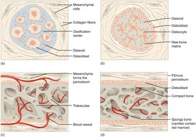
Stages of Endochondral Ossification
Endochondral ossification involves five key stages:
Formation of bone collar
Cavitation of hyaline cartilage
Invasion by periosteal bud and spongy bone formation
Formation of medullary cavity and secondary ossification centers
Ossification of epiphyses, with hyaline cartilage remaining in epiphyseal plates
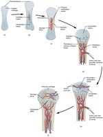
Long Bone Growth and Remodeling
Long bones grow in length and thickness through interstitial and appositional growth.
Interstitial growth: Cartilage grows and is replaced by bone, increasing length.
Appositional growth: Osteoblasts and osteoclasts remodel bone surfaces, increasing thickness.
Hormonal Regulation of Bone Growth
Bone growth is regulated by hormones.
Growth hormone: Stimulates epiphyseal plate activity.
Thyroid hormone: Modulates growth hormone activity.
Sex hormones: Promote growth spurts and induce epiphyseal plate closure at puberty.
Bone Deposition and Resorption
Bone is continuously deposited and resorbed to maintain strength and mineral homeostasis.
Deposition: Osteoblasts build bone; requires protein, vitamins, and minerals.
Resorption: Osteoclasts break down bone matrix; lysosomal enzymes digest organic matrix.
Control of Bone Remodeling
Bone remodeling is controlled by hormonal and mechanical factors.
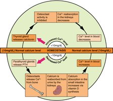
Hormonal mechanism: Maintains blood calcium homeostasis.
Mechanical forces: Stress and gravity influence bone structure.
Hormonal Control of Blood Calcium
Blood calcium levels are tightly regulated by hormones.
Low calcium: Parathyroid hormone (PTH) stimulates osteoclasts to release calcium from bone.
High calcium: Calcitonin from thyroid stimulates osteoblasts to deposit calcium in bone.
Response to Mechanical Stress
Wolff's law states that bone adapts to the forces placed upon it.
Observations: Bone thickens where stress is greatest; trabeculae align along stress lines; projections form where muscles attach.
Bone Fractures
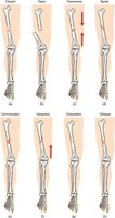
Bone fractures are classified by position, completeness, orientation, and skin penetration.
Nondisplaced: Ends retain normal position.
Displaced: Ends out of alignment.
Complete: Broken all the way through.
Incomplete: Not broken all the way through.
Linear: Parallel to long axis.
Common Types of Fractures
Transverse: Perpendicular to long axis.
Compound (open): Bone penetrates skin.
Simple (closed): Bone does not penetrate skin.
Comminuted: Bone fragments into pieces.
Spiral: Ragged break from twisting.
Depressed: Bone pressed inward.
Compression: Bone is crushed.
Greenstick: Incomplete fracture; common in children.
Bone Fracture Healing Stages
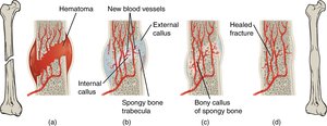
Bone healing occurs in four stages:
Hematoma formation
Fibrocartilaginous callus formation
Bony callus formation
Bone remodeling
Homeostatic Imbalances
Osteomalacia (Rickets)
Softening and weakening of bones due to inadequate mineralization, often from calcium or vitamin D deficiency. Symptoms include pain and deformities.
Osteoporosis
Loss of bone mass, common in elderly, especially in spine and femur. Risk factors include lack of estrogen, calcium, vitamin D, immobility, and certain diseases.
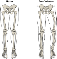
Paget's Disease
Excessive bone formation and breakdown, leading to weakened, spotty bone, often in spine, pelvis, femur, or skull. Cause is unknown.
Developmental Aspects of Bones
Bone development begins with mesoderm-derived mesenchymal cells forming the embryonic skeleton. Ossification follows a predictable timetable, with most bones ossified at birth except epiphyses. Bone mass peaks by age 25 and decreases with age, influenced by genetics and environment.
Bone Type | Location | Function |
|---|---|---|
Long | Limbs | Movement, support |
Short | Wrist, ankle | Stability, support |
Flat | Sternum, skull | Protection |
Irregular | Vertebrae, pelvis | Protection, support |
Sesamoid | Patella | Reduce friction |
Additional info: Table inferred for bone classification and function.
