 Indietro
Bones and Skeletal Tissues: Structure, Function, and Development
Guida di studio - Note intelligenti
 Appunti personalizzati basati sui tuoi materiali, ampliati con definizioni chiave, esempi e contesto.
Bones and Skeletal Tissues
Introduction
The human skeleton is a dynamic organ system composed of bones and cartilages. It provides structural support, protection, movement, and serves as a reservoir for minerals and site for blood cell formation. Understanding the structure, types, and functions of bones and skeletal tissues is fundamental in Anatomy & Physiology.
Skeletal Cartilages
General Structure and Function
Cartilage is a resilient, flexible connective tissue found in areas requiring support and flexibility.
It is avascular and lacks nerves, consisting mainly of water, which provides resilience.
Surrounded by the perichondrium, a dense irregular connective tissue that supplies nutrients and resists outward expansion.
Cartilage cells, called chondrocytes, are located in lacunae within a jelly-like extracellular matrix (ECM).
Types of Cartilage
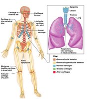
Hyaline Cartilage: Most abundant; provides support, flexibility, and resilience. Found in articular surfaces, costal cartilages, respiratory structures, and the nose.
Elastic Cartilage: Contains elastic fibers; found in the external ear and epiglottis. Withstands repeated bending.
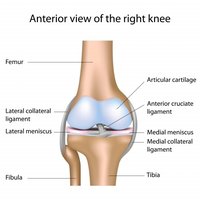
Fibrocartilage: Contains thick collagen fibers; highly compressible and strong. Found in intervertebral discs, menisci of the knee, and pubic symphysis.
Locations of Cartilage Types
Hyaline: Embryonic skeleton, articular cartilage of joints, costal cartilage, nose, larynx, trachea.
Elastic: Ear (pinna), epiglottis.
Fibrocartilage: Meniscus of knee, pubic symphysis, intervertebral discs.
Growth of Cartilage
Appositional growth: New matrix is secreted on the surface by chondroblasts in the perichondrium.
Interstitial growth: Chondrocytes within lacunae divide and secrete new matrix, expanding cartilage from within.
Calcification of cartilage can occur during bone growth or aging, but calcified cartilage is not bone.
Bones of the Skeleton
Axial vs. Appendicular Skeleton
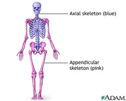
Axial skeleton: Forms the long axis of the body (skull, vertebral column, rib cage); functions in protection, support, and carrying body parts.
Appendicular skeleton: Bones of the limbs and girdles; involved in movement and manipulation of the environment.
Classification of Bones
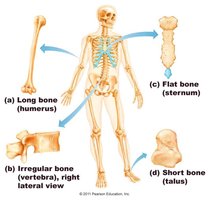
Long bones: Longer than wide (e.g., femur, humerus, phalanges).
Short bones: Cube-shaped (e.g., wrist and ankle bones).
Flat bones: Thin, flattened, often curved (e.g., sternum, scapulae, ribs, skull bones).
Irregular bones: Complicated shapes (e.g., vertebrae, hip bones).
Functions of Bones
Support: Framework for the body and soft organs.
Protection: Encloses vital organs (brain, spinal cord, thoracic organs).
Movement: Muscles attach to bones, using them as levers.
Mineral storage: Reservoir for calcium and phosphate.
Blood cell formation: Hematopoiesis in marrow cavities.
Triglyceride storage: Fat storage in bone cavities.
Hormone production: Osteocalcin regulates metabolism.
Bone Structure
Gross Anatomy
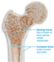
Compact bone: Dense outer layer; smooth and solid.
Spongy bone: Honeycomb of trabeculae filled with marrow.
Structure of Long Bones
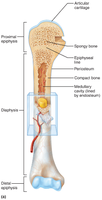
Diaphysis: Shaft; compact bone surrounds medullary cavity (contains yellow marrow in adults).
Epiphyses: Bone ends; compact bone exterior, spongy bone interior, articular cartilage covers joint surfaces.
Epiphyseal line: Remnant of growth plate between diaphysis and epiphysis.
Membranes: Periosteum (outer double-layered membrane) and endosteum (lines internal surfaces).
Structure of Short, Irregular, and Flat Bones
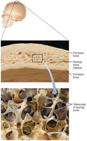
Thin plates of spongy bone (diploë) covered by compact bone.
No defined marrow cavity; marrow is scattered throughout spongy bone.
Periosteum covers outside; endosteum covers inside.
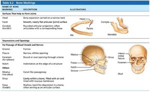
Bone Markings
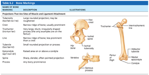
Bone markings are anatomical features that serve as sites for muscle, ligament, and tendon attachment, joint formation, or passageways for blood vessels and nerves.
Name of Bone Marking | Description | Illustration |
|---|---|---|
Crest | Narrow ridge of bone; usually prominent | Hip bone crest |
Condyle | Rounded articular projection | Mandible condyle |
Foramen | Round or oval opening through a bone | Skull foramen |
Fossa | Shallow, basinlike depression | Skull fossa |
Meatus | Canal-like passageway | Skull meatus |
Process | Any bony prominence | Vertebra process |
Microscopic Anatomy of Bone
Bone Cells
Osteoprogenitor cells: Stem cells in periosteum and endosteum; differentiate into osteoblasts or bone-lining cells.
Osteoblasts: Bone-forming cells; secrete osteoid (organic bone matrix).
Osteocytes: Mature bone cells in lacunae; maintain bone matrix and act as stress sensors.
Bone-lining cells: Flat cells on bone surfaces; help maintain matrix.
Osteoclasts: Multinucleate cells that resorb bone; derived from hematopoietic stem cells.
Compact Bone Structure
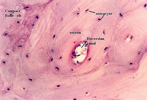
Osteon (Haversian system): Structural unit; concentric lamellae around a central canal.
Lamellae: Collagen fibers in different directions; resist twisting.
Canals and canaliculi: Central canal contains blood vessels and nerves; canaliculi connect osteocytes for nutrient/waste exchange.
Lacunae: Small cavities containing osteocytes.
Interstitial and circumferential lamellae: Fill gaps and encircle the diaphysis, respectively.
Spongy Bone Structure
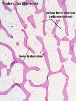
Composed of trabeculae aligned along lines of stress.
No osteons; contains irregularly arranged lamellae, osteocytes, and canaliculi.
Capillaries in endosteum supply nutrients.
Chemical Composition of Bone
Organic components: Cells and osteoid (collagen fibers and ground substance); provide tensile strength and flexibility.
Inorganic components: Hydroxyapatites (mineral salts, mainly calcium phosphate); provide hardness and resistance to compression.
Bone Development (Osteogenesis/Ossification)
Overview
Osteogenesis is the process of bone tissue formation.
Occurs during embryonic development, postnatal growth, and bone remodeling/repair in adults.
Formation of the Bony Skeleton
Endochondral ossification: Bone forms by replacing hyaline cartilage; forms most bones below the skull except clavicles.
Intramembranous ossification: Bone develops from fibrous membranes; forms flat bones of skull and clavicles.
Postnatal Bone Growth
Interstitial growth: Lengthening at the epiphyseal plate.
Appositional growth: Increase in bone thickness.
Growth regulated by growth hormone, thyroid hormone, and sex hormones.
Bone Remodeling and Repair
Bone Remodeling
Continuous process involving bone deposit (by osteoblasts) and resorption (by osteoclasts).
Regulated by hormonal controls (parathyroid hormone, calcitonin) and response to mechanical stress (Wolff's law).
Low blood Ca2+: Parathyroid hormone stimulates osteoclasts to release Ca2+ from bone.
High blood Ca2+: Calcitonin (minor effect) may stimulate Ca2+ deposition in bone.
Bone Repair
Fracture repair involves four stages: hematoma formation, fibrocartilaginous callus formation, bony callus formation, and bone remodeling.
Bone Disorders
Osteomalacia and Rickets
Caused by vitamin D deficiency or insufficient calcium.
Osteomalacia: Poorly mineralized, soft bones in adults.
Rickets: Osteomalacia in children; results in bone deformities.
Osteoporosis
Bone resorption exceeds deposit; bone mass declines.
Common in postmenopausal women; risk factors include low exercise, poor diet, smoking, genetics, and certain diseases.
Prevention: Weight-bearing exercise, adequate calcium and vitamin D, medications that inhibit osteoclasts.
Paget’s Disease
Excessive, disorganized bone remodeling; high ratio of spongy to compact bone.
Common in spine, pelvis, femur, and skull; cause unknown.
Treatment includes calcitonin and bisphosphonates.
Additional info: This guide covers the essential structure, function, and pathology of bones and skeletal tissues, integrating key diagrams and tables for visual reinforcement.
