 Indietro
Peripheral Nervous System: Spinal and Cranial Nerves, Plexuses, and Clinical Correlates
Guida di studio - Note intelligenti
 Appunti personalizzati basati sui tuoi materiali, ampliati con definizioni chiave, esempi e contesto.
Peripheral Nervous System (PNS) Overview
Introduction to the PNS
The Peripheral Nervous System (PNS) connects the Central Nervous System (CNS) to the rest of the body, enabling communication between the brain, spinal cord, and peripheral tissues. It is divided into sensory (afferent) and motor (efferent) divisions, each with somatic and visceral components.
Sensory Division: Transmits sensory information from receptors to the CNS.
Motor Division: Transmits motor commands from the CNS to effectors (muscles and glands).
Somatic refers to structures of the body wall (skin, skeletal muscle), while visceral refers to internal organs.
PNS Functional Overview
Sensory (Afferent) Pathways: Detect stimuli and relay signals to the CNS.
Motor (Efferent) Pathways: Carry commands from the CNS to effectors.
Somatic Motor: Voluntary control of skeletal muscles.
Visceral Motor (Autonomic Nervous System, ANS): Involuntary control of smooth muscle, cardiac muscle, and glands.
The ANS is further divided into:
Sympathetic Division: Prepares the body for 'fight or flight' responses.
Parasympathetic Division: Maintains 'rest and digest' functions.
Classification of Peripheral Nerves
Functional Classification
Mixed Nerves: Contain both sensory and motor fibers; most common type.
Sensory Nerves: Carry only sensory information to the CNS.
Motor Nerves: Carry primarily motor commands, but may contain some sensory fibers for feedback.
Anatomical Classification
Spinal Nerves: Originate from the spinal cord; innervate structures below the head and neck.
Cranial Nerves: Originate from the brain; innervate structures of the head and neck.
Spinal Nerves and Plexuses
Structure of Spinal Nerves
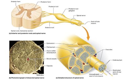
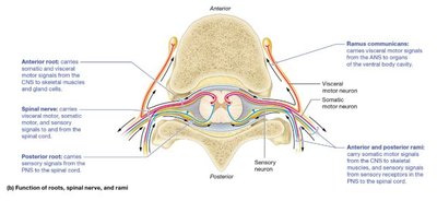
Spinal nerves are formed by the fusion of anterior (motor) and posterior (sensory) roots. Each nerve quickly splits into posterior and anterior rami, which innervate different regions of the body.
Posterior Root: Contains sensory fibers entering the spinal cord.
Anterior Root: Contains motor fibers exiting the spinal cord.
Posterior Ramus: Innervates muscles and skin of the back.
Anterior Ramus: Innervates anterior and lateral trunk, limbs; forms nerve plexuses.
Rami Communicantes: Carry autonomic (visceral motor) fibers to and from the sympathetic trunk.
Detailed Structure of a Spinal Nerve
Epineurium: Outermost connective tissue covering.
Perineurium: Surrounds bundles (fascicles) of axons.
Endoneurium: Surrounds individual axons.
Spinal Nerve Plexuses
Plexuses are networks of intersecting nerves formed by the anterior rami of spinal nerves. They provide redundancy and protection against injury.
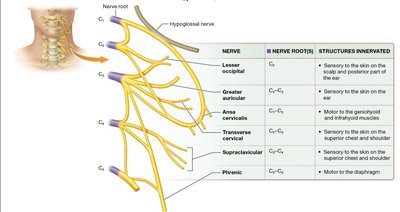
Cervical Plexus (C1–C4): Innervates neck, diaphragm (via phrenic nerve).
Brachial Plexus (C5–T1): Innervates upper limb.
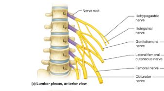
Lumbar Plexus (L1–L4): Innervates anterior and medial thigh.
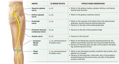
Sacral Plexus (L4–S4): Innervates posterior thigh, most of lower leg and foot.
Cervical Plexus
Lesser Occipital (C2): Sensory to scalp and posterior ear.
Greater Auricular (C2–C3): Sensory to ear and parotid region.
Transverse Cervical (C2–C3): Sensory to anterior neck.
Supraclavicular (C3–C4): Sensory to shoulder and chest.
Phrenic (C3–C5): Motor to diaphragm ("3, 4, 5 to stay alive").
Brachial Plexus
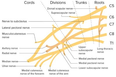
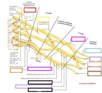
The brachial plexus is organized into roots, trunks, divisions, cords, and terminal branches. It innervates the shoulder, arm, forearm, and hand.
Axillary Nerve: Deltoid, teres minor, skin over deltoid.
Radial Nerve: Posterior arm/forearm, triceps brachii, extensors.
Musculocutaneous Nerve: Anterior arm, biceps brachii, lateral forearm skin.
Median Nerve: Forearm flexors, some hand muscles, lateral palm.
Ulnar Nerve: Forearm flexors not innervated by median, most intrinsic hand muscles, medial hand.
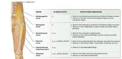
Lumbar Plexus
Iliohypogastric (L1): Motor to abdominal muscles, sensory to lower abdomen.
Ilioinguinal (L1): Motor to abdominal muscles, sensory to groin.
Genitofemoral (L1–L2): Sensory to anterior thigh, scrotum/labia.
Femoral (L2–L4): Motor to quadriceps, sensory to anterior/medial thigh and leg.
Obturator (L2–L4): Motor to thigh adductors, sensory to medial thigh.
Sacral Plexus
Superior Gluteal (L4–S1): Motor to gluteus medius/minimus.
Inferior Gluteal (L5–S2): Motor to gluteus maximus.
Sciatic Nerve (L4–S3): Largest nerve, splits into tibial and common fibular nerves.
Tibial Nerve: Posterior thigh/leg, plantar foot.
Common Fibular (Peroneal) Nerve: Anterior/lateral leg, dorsum of foot.
Cranial Nerves
Overview and Classification
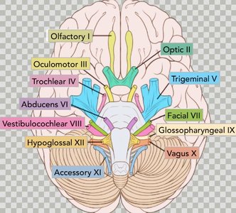
There are 12 pairs of cranial nerves, each with a specific name, number (Roman numeral), origin, and function. They may be sensory, motor, or mixed.
Sensory Only: Olfactory (I), Optic (II), Vestibulocochlear (VIII)
Motor Only: Oculomotor (III), Trochlear (IV), Abducens (VI), Accessory (XI), Hypoglossal (XII)
Mixed: Trigeminal (V), Facial (VII), Glossopharyngeal (IX), Vagus (X)
Key Cranial Nerves and Functions
Olfactory (I): Smell (sensory)
Optic (II): Vision (sensory)
Oculomotor (III): Eye movement, pupil constriction (motor)
Trochlear (IV): Eye movement (motor)
Trigeminal (V): Facial sensation, mastication (mixed)
Abducens (VI): Eye movement (motor)
Facial (VII): Facial expression, taste (anterior 2/3 tongue), glands (mixed)
Vestibulocochlear (VIII): Hearing, balance (sensory)
Glossopharyngeal (IX): Taste (posterior 1/3 tongue), swallowing, salivation (mixed)
Vagus (X): Parasympathetic to thoracic/abdominal organs, taste, swallowing (mixed)
Accessory (XI): Head/shoulder movement, speech (motor)
Hypoglossal (XII): Tongue movement (motor)
Clinical Correlates
Trigeminal Neuralgia
Severe, brief facial pain due to trigeminal nerve irritation or compression.
Triggers: chewing, touch, vibration, breeze.
Treatment: medications to reduce abnormal nerve transmission; painkillers often ineffective.
Bell's Palsy
Facial nerve (VII) dysfunction, often due to viral infection, trauma, or tumor.
Symptoms: facial muscle weakness/paralysis, difficulty blinking, altered taste, salivation issues.
Treatment: anti-inflammatory/antiviral medications, physical therapy; usually temporary.
Practice and Application
Label diagrams of the brachial plexus and identify major nerves and their regions of innervation.
Practice identifying cranial nerves by name, number, and function.
Summary Table: Major Nerve Plexuses and Key Nerves
Plexus | Major Nerves | Regions Innervated |
|---|---|---|
Cervical (C1–C4) | Phrenic, Lesser Occipital, Greater Auricular, Transverse Cervical, Supraclavicular | Neck, diaphragm, shoulder, chest |
Brachial (C5–T1) | Axillary, Radial, Musculocutaneous, Median, Ulnar | Shoulder, arm, forearm, hand |
Lumbar (L1–L4) | Femoral, Obturator, Iliohypogastric, Ilioinguinal, Genitofemoral | Anterior/medial thigh, lower abdomen, groin |
Sacral (L4–S4) | Sciatic, Tibial, Common Fibular, Superior/Inferior Gluteal | Posterior thigh, leg, foot, gluteal region |
Additional info: The PNS is essential for voluntary and involuntary control of body functions, and its organization into plexuses provides redundancy and protection against injury. Understanding the structure and function of cranial and spinal nerves is critical for diagnosing and treating neurological disorders.
