 Indietro
The Cell: Structure, Function, and Membrane Transport
Guida di studio - Note intelligenti
 Appunti personalizzati basati sui tuoi materiali, ampliati con definizioni chiave, esempi e contesto.
The Cell: General Structure
Overview of Cell Types
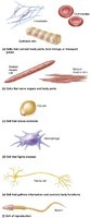
The human body contains approximately 200 distinct types of cells, each with unique shapes, sizes, and functions. Cells are the basic structural and functional units of life, and their diversity enables the body to perform a wide range of physiological tasks.
Key Point 1: Cells vary in morphology, including spherical, elongated, and irregular shapes.
Key Point 2: Specialized cells perform specific functions, such as nerve transmission, muscle contraction, and immune defense.
Example: Red blood cells transport oxygen, neurons transmit electrical signals, and muscle cells contract to produce movement.
Major Cell Components
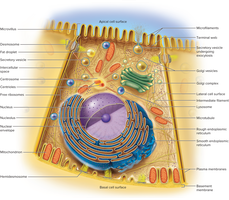
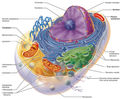
All cells share three fundamental components: the plasma membrane, cytoplasm, and nucleus. These structures are essential for maintaining cellular integrity and regulating internal processes.
Plasma membrane: The boundary separating the cell's internal environment from the external environment.
Cytoplasm: The fluid and organelles within the cell, excluding the nucleus.
Nucleus: The control center containing DNA and directing cellular activities.
The Cell Surface
Functions of the Cell Surface
The cell surface is a dynamic interface where essential processes occur, including communication, adhesion, and transport. The plasma membrane is the primary structure at the cell surface.
Key Point 1: The cell surface binds signaling molecules, such as hormones, to stimulate cellular activity.
Key Point 2: It enables attachment between cells and regulates the transport of materials in and out of the cell.
Plasma Membrane Composition
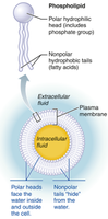
The plasma membrane is a selectively permeable barrier composed mainly of phospholipids, cholesterol, and glycolipids. Its fluid nature allows for flexibility and adaptability.
Phospholipids: Form a bilayer with hydrophilic heads facing outward and hydrophobic tails inward.
Cholesterol: Stabilizes membrane fluidity.
Glycolipids: Contribute to cell recognition and signaling.
Membrane Proteins
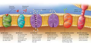
Membrane proteins are critical for cell function and vary by cell type. They are classified as peripheral (on the inner surface) or integral (spanning the membrane).
Channels: Allow selective passage of solutes.
Gated channels: Open or close in response to stimuli.
Receptors: Bind chemical messengers.
Enzymes: Catalyze reactions at the membrane.
Cell identifier markers: Distinguish self from foreign cells.
Cell adhesion molecules: Bind cells together.
Cell Surface Extensions
Cells may possess extensions that increase surface area or facilitate movement.
Microvilli: Increase surface area for absorption.
Cilia: Move substances across the cell surface.
Flagella: Propel cells (e.g., sperm).
Pseudopods: Enable cell movement and engulfment.
Cell Junctions
Cell junctions are protein complexes that link cells to each other and the extracellular environment, enabling communication, resistance to stress, and coordinated growth.
Tight junctions: Seal cells together, preventing passage of substances.
Desmosomes: Provide mechanical strength by anchoring cells.
Gap junctions: Allow direct communication between cells via channels.
Membrane Transport
Overview of Membrane Transport
Substances move across the plasma membrane by passive or active mechanisms. Passive transport requires no energy, while active transport uses ATP.
Passive transport: Includes filtration, simple diffusion, and osmosis.
Active transport: Includes carrier-mediated and vesicular transport.
Filtration
Filtration is the movement of fluid and small particles through a selectively permeable membrane driven by physical pressure.
Key Point: Allows passage of water and small solutes, but blocks larger particles.
Simple Diffusion
Simple diffusion is the movement of particles down their concentration gradient without energy input. Small, non-polar solutes diffuse through the lipid regions of the plasma membrane.
Examples: Oxygen, carbon dioxide, steroid hormones.
Osmosis
Osmosis is the net movement of water across a selectively permeable membrane, from areas of low solute concentration to high solute concentration.
Key Point: Osmosis is vital for maintaining cell volume and internal pressure.
Tonicity: The ability of a solution to affect intracellular pressure and volume, determined by solute concentration.
Isotonic: Equal solute concentration inside and outside the cell.
Hypertonic: Higher solute concentration outside; cell shrivels.
Hypotonic: Lower solute concentration outside; cell swells.
Carrier-Mediated Transport
Carrier-mediated transport involves proteins that facilitate the movement of solutes across the membrane.
Facilitated diffusion: Solute moves down its concentration gradient with the help of a carrier; no energy required.
Active transport: Solute moves against its concentration gradient using a carrier protein and ATP.
Vesicular Transport
Vesicular transport moves particles in bubble-like vesicles and requires ATP. It includes endocytosis (bringing matter into cells) and exocytosis (expelling matter from cells).
Phagocytosis: Cell engulfs particles using pseudopods.
Pinocytosis: Cell takes in extracellular fluid.
Receptor-mediated endocytosis: Selective uptake of specific molecules.
Exocytosis: Discharge of material from the cell.
Cell Interior: Organelles
Overview of Organelles
Organelles are specialized structures within cells that perform distinct functions, analogous to organs in the body. They compartmentalize cellular processes and maintain order.
Cytoskeleton
The cytoskeleton is a network of protein filaments that provides structural support, facilitates movement, and organizes cell contents.
Microfilaments: Thin filaments involved in cell movement.
Intermediate filaments: Provide mechanical strength.
Microtubules: Hollow tubes for intracellular transport and cell division.
Nucleus
The nucleus is the largest organelle, surrounded by a nuclear envelope, and contains chromosomes and the nucleolus. It directs cellular activities and stores genetic information.
Nucleolus: Site of ribosome assembly.
Endoplasmic Reticulum (ER)
The ER is a network of membranes with two regions: rough (RER) and smooth (SER).
Rough ER: Covered in ribosomes; synthesizes proteins for secretion.
Smooth ER: Lacks ribosomes; synthesizes lipids and detoxifies substances.
Ribosomes
Ribosomes are composed of proteins and RNA. They read genetic messages and assemble amino acids into proteins.
Free ribosomes: Make proteins for use within the cell.
Bound ribosomes: Make proteins for export.
Golgi Complex
The Golgi complex synthesizes carbohydrates and modifies proteins. It packages proteins into vesicles for transport or secretion.
Lysosomes
Lysosomes are membrane-bound enzyme packages that digest cellular debris and participate in programmed cell death (apoptosis).
Clinical Example: Tay-Sachs disease results from defective lysosomal enzymes, leading to neurodegeneration.
Peroxisomes
Peroxisomes contain enzymes that detoxify harmful substances and break down fatty acids. They neutralize free radicals and protect the cell.
Mitochondria
Mitochondria are the "powerhouse" of the cell, responsible for ATP synthesis. They have a double membrane, inner folds (cristae), and their own DNA.
Key Point: ATP is the cell's energy currency, produced by mitochondria.
Centrioles
Centrioles are cylindrical structures that form mitotic spindles during cell division. The centrosome is a pair of centrioles near the nucleus.
The Cell Life Cycle
Phases of the Cell Cycle
Cells progress through a life cycle consisting of interphase and mitosis. Interphase includes G1, S, and G2 phases, while mitosis divides the nucleus and cytoplasm.
G1: Cell grows and synthesizes proteins.
S: DNA replication occurs.
G2: Cell prepares for division.
Mitosis: Nucleus divides, followed by cytokinesis.
Mitosis Stages
Mitosis is divided into four stages: prophase, metaphase, anaphase, and telophase.
Prophase: Chromosomes condense, nuclear envelope disintegrates, spindle fibers form.
Metaphase: Chromosomes align at the cell center.
Anaphase: Sister chromatids separate and migrate to opposite poles.
Telophase: Chromatids cluster, new nuclear envelopes form, cytokinesis begins.
Summary Table: Cell Organelles and Functions
Organelle | Function |
|---|---|
Nucleus | Stores genetic material, directs cell activities |
Ribosomes | Protein synthesis |
Rough ER | Synthesizes proteins for secretion |
Smooth ER | Synthesizes lipids, detoxifies substances |
Golgi Complex | Modifies, packages, and sorts proteins |
Lysosomes | Digest cellular debris, apoptosis |
Peroxisomes | Detoxify substances, break down fatty acids |
Mitochondria | ATP synthesis |
Centrioles | Form mitotic spindles |
Key Formula: Osmosis
The rate of osmosis can be described by Fick's law:
J: Flux (rate of movement)
D: Diffusion coefficient
\frac{dC}{dx}: Concentration gradient
Key Formula: Active Transport
Active transport requires energy input:
ATP: Adenosine triphosphate
ADP: Adenosine diphosphate
Pi: Inorganic phosphate
