 Indietro
The Cell: Structure, Function, and Membrane Transport
Guida di studio - Note intelligenti
 Appunti personalizzati basati sui tuoi materiali, ampliati con definizioni chiave, esempi e contesto.
The Cell
General Structure of Cells
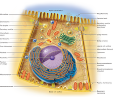
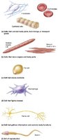
The cell is the fundamental unit of life in the human body, with approximately 200 distinct types varying in shape, size, and function. Each cell type is specialized to perform unique tasks essential for bodily function.
Cell Components:
Plasma membrane: Boundary separating the cell from its environment.
Cytoplasm: Internal fluid containing organelles.
Nucleus: Control center containing DNA.
Variety of Cell Types: Cells can be flat, spherical, elongated, or branched, reflecting their specialized roles.
Cell Interior: Organelles
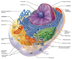
Organelles are specialized structures within cells, each performing distinct metabolic functions. Compartmentalization allows cells to maintain order and efficiency.
Nucleus: Largest organelle, surrounded by a nuclear envelope, contains chromosomes and nucleolus.
Endoplasmic Reticulum (ER):
Rough ER: Covered in ribosomes, synthesizes proteins for secretion.
Smooth ER: Lacks ribosomes, synthesizes lipids and detoxifies substances.
Ribosomes: Sites of protein synthesis, either free in cytoplasm or bound to ER.
Golgi Complex: Modifies, sorts, and packages proteins and carbohydrates.
Lysosomes: Membrane-bound enzyme packages for digestion and apoptosis.
Peroxisomes: Detoxify harmful substances and break down fatty acids.
Mitochondria: Powerhouse of the cell, site of ATP synthesis, contains its own DNA.
Centrioles: Organize microtubules during cell division.
The Cell Surface
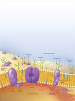
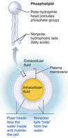
The cell surface is a dynamic interface where communication, attachment, and transport occur. It is composed mainly of the plasma membrane, which separates intracellular fluid (ICF) from extracellular fluid (ECF).
Plasma Membrane:
Phospholipid bilayer (75%) provides fluidity and selective permeability.
Cholesterol (20%) stabilizes membrane structure.
Glycolipids (5%) contribute to cell recognition.
Membrane Proteins:
Peripheral proteins: Located on the inner surface.
Integral proteins: Span the membrane, forming channels and receptors.
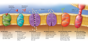
Membrane Proteins and Their Functions
Membrane proteins are critical for cell function, enabling transport, communication, and identification.
Channel proteins: Form pores for selective solute passage.
Gated channels: Open or close in response to stimuli.
Receptors: Bind chemical messengers (e.g., hormones).
Enzymes: Catalyze reactions at the membrane surface.
Cell identifier markers: Distinguish self from foreign cells.
Cell adhesion molecules: Bind cells together.
Cell Surface Extensions
Cells may have surface extensions that increase surface area or aid in movement.
Microvilli: Increase surface area for absorption.
Cilia: Move substances across cell surfaces.
Flagella: Propel cells (e.g., sperm).
Pseudopods: Temporary extensions for movement and engulfing particles.
Cell Junctions
Cell junctions are protein complexes that link cells to each other and to the extracellular matrix, enabling communication, resistance to stress, and coordinated growth.
Tight junctions: Seal cells together, preventing passage between them.
Desmosomes: Rivet-like junctions providing mechanical strength.
Gap junctions: Channels allowing direct communication and passage of ions and small molecules.
Membrane Transport
Substances move across the plasma membrane by passive or active mechanisms, maintaining cellular homeostasis.
Passive Transport: No energy required. Includes filtration, simple diffusion, and osmosis.
Active Transport: Requires ATP. Includes carrier-mediated transport and vesicular transport.
Filtration
Filtration uses physical pressure to drive fluid through a selectively permeable membrane, allowing water and small particles to pass while blocking larger ones.
Simple Diffusion
Simple diffusion is the movement of particles down their concentration gradient without energy input. Small, non-polar solutes (e.g., oxygen, carbon dioxide, steroid hormones) diffuse through the lipid bilayer.
Osmosis
Osmosis is the net movement of water across a selectively permeable membrane, from areas of low solute concentration to high solute concentration. It is essential for maintaining cell volume and pressure.
Tonicity:
Isotonic: Equal solute concentration inside and outside the cell.
Hypertonic: Higher solute concentration outside; cell shrivels.
Hypotonic: Lower solute concentration outside; cell swells.
Carrier-Mediated Transport
Facilitated diffusion: Carrier proteins help solutes move down their concentration gradient without energy.
Active transport: Carrier proteins use ATP to move solutes against their concentration gradient.
Vesicular Transport
Endocytosis: Bringing matter into the cell via vesicles.
Exocytosis: Expelling matter from the cell via vesicles.
Phagocytosis: Cell engulfs particles (e.g., by immune cells).
Pinocytosis: Cell takes in fluid.
Receptor-mediated endocytosis: Selective uptake of specific molecules.
Cell Interior: Cytoskeleton
The cytoskeleton is a network of protein filaments providing structural support, movement, and organization within the cell.
Microfilaments
Intermediate filaments
Microtubules
The Cell Life Cycle
Cells undergo a life cycle consisting of interphase and mitosis, enabling growth, DNA replication, and division.
Interphase:
G1: Cell grows and synthesizes proteins.
S: DNA replication.
G2: Preparation for division.
Mitosis:
Prophase: Chromosomes condense, spindle fibers form.
Metaphase: Chromosomes align at the center.
Anaphase: Chromatids separate.
Telophase: Chromatids cluster, nuclear envelope reforms, cytokinesis occurs.
Clinical Example: Tay-Sachs Disease
Tay-Sachs disease is a genetic disorder caused by a deficiency in a lysosomal enzyme, leading to accumulation of glycolipids in nerve cells. Symptoms include developmental regression, blindness, deafness, seizures, and early death.
Key Terms and Definitions
Amphipathic: Molecule with both hydrophilic and hydrophobic regions (e.g., phospholipids).
ATP: Adenosine triphosphate, the energy currency of the cell.
Apoptosis: Programmed cell death.
Homeostasis: Maintenance of stable internal conditions.
Summary Table: Membrane Transport Mechanisms
Transport Type | Energy Required | Direction | Example |
|---|---|---|---|
Filtration | No | Down pressure gradient | Kidney filtration |
Simple Diffusion | No | Down concentration gradient | Oxygen diffusion |
Osmosis | No | Water down concentration gradient | Cell volume regulation |
Facilitated Diffusion | No | Down concentration gradient | Glucose transport |
Active Transport | Yes (ATP) | Against concentration gradient | Sodium-potassium pump |
Vesicular Transport | Yes (ATP) | Variable | Endocytosis, exocytosis |
Key Equations
Osmosis: Water movement is driven by solute concentration differences.
Simple Diffusion: Rate of diffusion is proportional to concentration gradient.
Example Application
In the kidneys, filtration allows waste products to be removed from the blood while retaining larger molecules like proteins. In neurons, gap junctions enable rapid electrical communication.
