 Indietro
The Skeletal System: Bones and Skeletal Tissues
Guida di studio - Note intelligenti
 Appunti personalizzati basati sui tuoi materiali, ampliati con definizioni chiave, esempi e contesto.
Introduction to the Skeletal System
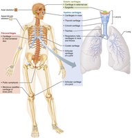
The skeletal system is the living framework of bones, cartilage, and connective tissues that supports the human body. An adult human skeleton typically contains 206 bones, which provide structure, protection, and facilitate movement.
The Skeletal System
Parts of the Skeletal System
Bones (skeleton): The rigid organs that form the skeleton.
Joints: Articulations where two or more bones meet.
Cartilages: Flexible connective tissue found in joints, ear, nose, and respiratory tract.
Ligaments: Bands of dense connective tissue connecting bones at joints.
Subdivisions of the Skeleton
Axial skeleton: Forms the long axis of the body (skull, vertebral column, rib cage).
Appendicular skeleton: Bones of the limbs and girdles (shoulder and pelvic girdles).
The Skeletal System: Functions
The skeleton serves several vital functions:
Support: Provides a structural framework for the body.
Protection: Shields vital organs (e.g., skull protects the brain, rib cage protects thoracic organs).
Movement: Acts as levers for muscles to produce movement.
Storage: Stores minerals (calcium, phosphorus) and fats (in marrow).
Blood cell formation (hematopoiesis): Occurs in red marrow cavities.
Hormone production: Bones produce osteocalcin, which helps regulate bone formation and metabolism.
The Skeletal System: Classification of Bones
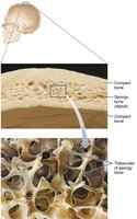
Types of Osseous Tissue
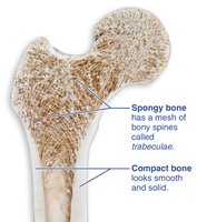
Compact bone: Dense, smooth, and homogeneous tissue.
Spongy bone: Composed of small needlelike pieces (trabeculae) and many open spaces.
Classification by Shape
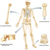
Long bones: Longer than wide, shaft with enlarged ends (e.g., femur, humerus).
Flat bones: Thin, flattened, usually curved (e.g., skull, ribs, sternum).
Short bones: Cube-shaped, mostly spongy bone (e.g., carpals, tarsals, patella).
Irregular bones: Complex shapes that do not fit other categories (e.g., vertebrae, hip bones).
Bone Structure
Typical Long Bone Anatomy
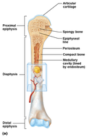
Diaphysis: Shaft, composed of compact bone.
Epiphysis: Ends, mostly spongy bone enclosed by compact bone.
Periosteum: Outer fibrous membrane covering diaphysis, secured by Sharpey's fibers.
Articular cartilage: Hyaline cartilage covering epiphyses, reduces friction at joints.
Epiphyseal plate/line: Growth plate of hyaline cartilage (plate in youth, line in adults).
Endosteum: Membrane lining the medullary cavity.
Medullary cavity: Central cavity containing yellow marrow (fat) in adults, red marrow in children.
Microscopic Anatomy of Bone
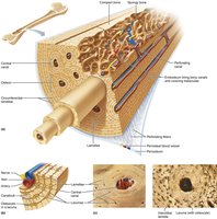
Spongy bone: Trabeculae form a network filled with marrow, blood vessels, and nerves.
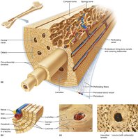
Compact bone: Organized into osteons (Haversian systems) with central canals, lamellae, lacunae, and canaliculi.
Osteocytes: Mature bone cells in lacunae.
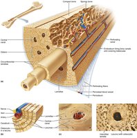
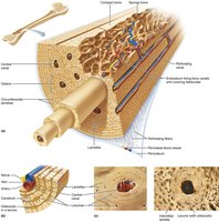
Lamellae: Concentric rings of bone matrix around central canal.
Canaliculi: Tiny canals connecting lacunae to each other and to the central canal.
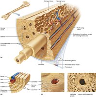
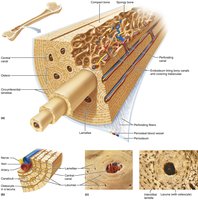
Central (Haversian) canal: Runs lengthwise, contains blood vessels and nerves.
Perforating (Volkmann's) canal: Runs perpendicular, connects blood and nerve supply of periosteum to central canals.
Cells of Bone Tissue
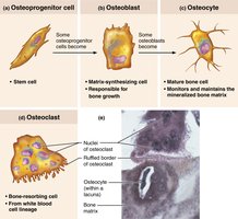
Osteogenic cells: Stem cells that differentiate into osteoblasts.
Osteoblasts: Bone-forming cells responsible for bone growth.
Osteocytes: Mature bone cells that maintain bone matrix.
Osteoclasts: Bone-resorbing cells derived from white blood cell lineage.
Bone Markings
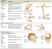
Bone markings are anatomical features on bones that serve as sites for muscle, tendon, and ligament attachment, or as passages for nerves and blood vessels.
Category | Marking | Description |
|---|---|---|
Projection | Tuberosity, Crest, Trochanter, Line, Tubercle, Epicondyle, Spine, Process, Head, Facet, Condyle | Sites of muscle and ligament attachment or articulation |
Depression/Opening | Groove, Fissure, Foramen, Notch, Meatus, Sinus, Fossa | Passages for nerves and blood vessels |
Bone Composition
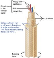
Organic components: Collagen fibers provide flexibility and tensile strength.
Inorganic components: Calcium salts (hydroxyapatite) provide hardness and resistance to compression.
Bone Formation, Growth, and Remodeling
Ossification (Bone Formation)
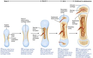
Endochondral ossification: Bone forms by replacing hyaline cartilage (most bones of the body).
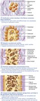
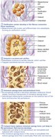
Intramembranous ossification: Bone develops from fibrous membranes (flat bones of skull, clavicle).
Bone Growth
Lengthwise growth: Occurs at the epiphyseal plate by cartilage growth and replacement by bone.
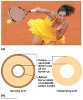
Appositional growth: Bones increase in diameter by addition of bone matrix to the outer surface by osteoblasts and removal from the inner surface by osteoclasts.
Hormonal regulation: Growth hormone and sex hormones regulate bone growth.
Bone Remodeling
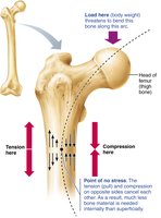
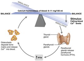
Bones are continuously remodeled in response to blood calcium levels and mechanical stress.
Parathyroid hormone (PTH): Increases blood calcium by stimulating osteoclasts to break down bone.
Calcitonin: Lowers blood calcium by stimulating osteoblasts to deposit bone matrix (not detailed in the provided text but relevant for context).
Bone Fractures
Types of Bone Fractures
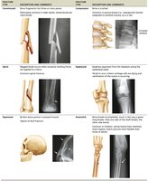
Closed (simple) fracture: Bone breaks but does not penetrate the skin.
Open (compound) fracture: Broken bone penetrates the skin.

Other types: Comminuted, spiral, depressed, compression, epiphyseal, greenstick.
Repair of Bone Fractures
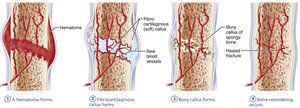
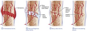
Bone repair involves four major stages:
Hematoma formation
Fibrocartilaginous callus formation
Bony callus formation
Bone remodeling
Developmental Aspects of the Skeleton
From Birth to Adulthood
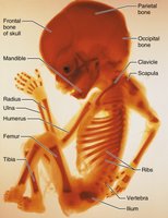
Fetal long bones are initially hyaline cartilage; flat bones form from fibrous membranes.
Fontanels (soft spots) in the fetal skull allow for brain growth and skull compression during birth.
Fontanels ossify by age 2.
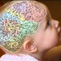
Skull reaches near-adult size by age 8 or 9; facial bones grow with tooth development and airway enlargement.
During puberty, the female pelvis broadens and the male skeleton becomes more robust.
Epiphyseal plates ossify by the end of adolescence.
Aging and the Skeleton
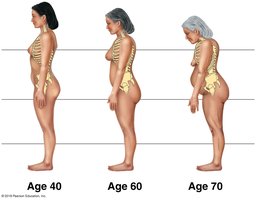
Bones lose density and become more brittle with age.
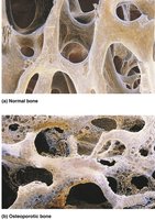
Osteoporosis: Bone-thinning disease common in postmenopausal women and elderly men, leading to increased fracture risk and kyphosis (dowager's hump).
Estrogen helps maintain bone density in females.
Homeostatic Imbalances
Osteomalacia: Poorly mineralized bones, resulting in soft, weak bones and pain upon bearing weight.
Rickets: Osteomalacia in children, causing bone deformities due to vitamin D or calcium deficiency.
Giantism: Oversecretion of growth hormone in childhood.
Short stature: Undersecretion of growth or thyroid hormone in childhood.
