Ch. 11 Cardiovascular System
Marieb - Essentials of Human Anatomy & Physiology 13th Edition
Marieb13th EditionEssentials of Human Anatomy & PhysiologyISBN: 9780135624340Non è quello che usi tu?Cambia libro di testo
Capitolo 11, Problema 20

Draw a diagram of the heart showing the three layers composing its wall and its four chambers. Label each. Show where the AV and semilunar valves are, and name them. Show and label all blood vessels entering and leaving the heart chambers.

Guida verificata passo dopo passo
1
Begin by drawing the outline of the heart. Divide it into four chambers: two upper chambers (atria) and two lower chambers (ventricles). Label the right atrium, left atrium, right ventricle, and left ventricle.
Next, illustrate the three layers of the heart wall. Start with the innermost layer (endocardium), followed by the middle muscular layer (myocardium), and finally the outermost layer (epicardium). Label each layer clearly.
Draw and label the atrioventricular (AV) valves: the tricuspid valve between the right atrium and right ventricle, and the mitral (bicuspid) valve between the left atrium and left ventricle. Then, add the semilunar valves: the pulmonary valve at the exit of the right ventricle and the aortic valve at the exit of the left ventricle.
Illustrate and label the major blood vessels entering and leaving the heart chambers. Include the superior and inferior vena cava entering the right atrium, the pulmonary arteries leaving the right ventricle, the pulmonary veins entering the left atrium, and the aorta leaving the left ventricle.
Ensure all components are clearly labeled and visually organized. Highlight the flow of blood through the heart by using arrows to indicate the direction of blood movement: from the vena cava to the right atrium, through the tricuspid valve to the right ventricle, out through the pulmonary valve to the pulmonary arteries, back via the pulmonary veins to the left atrium, through the mitral valve to the left ventricle, and out through the aortic valve to the aorta.

Risposta video verificata per un problema simile:

Questa soluzione video è stata consigliata dai nostri tutor come utile per risolvere questo problema.

Concetti chiave

Ecco i concetti essenziali che devi comprendere per rispondere correttamente alla domanda.

Heart Anatomy

The heart is a muscular organ divided into four chambers: the right atrium, right ventricle, left atrium, and left ventricle. Each chamber plays a crucial role in the circulation of blood, with the atria receiving blood and the ventricles pumping it out. Understanding the layout of these chambers is essential for accurately drawing and labeling the heart.
Video consigliato:
6:13
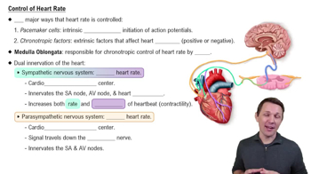
Control of Heart Rate

Heart Wall Layers

The heart wall consists of three layers: the epicardium (outer layer), myocardium (middle layer), and endocardium (inner layer). The epicardium provides a protective layer, the myocardium is responsible for the heart's contraction, and the endocardium lines the chambers and valves. Recognizing these layers is important for a comprehensive diagram of the heart.
Video consigliato:
Percorso guidato
02:31
Epidermal Layers: Stratum Lucidum (Clear Layer)

Valves and Blood Flow

The heart contains four main valves: the tricuspid and mitral (AV valves) and the pulmonary and aortic (semilunar valves). These valves ensure unidirectional blood flow through the heart and prevent backflow. Identifying their locations and functions is crucial for understanding how blood circulates through the heart and the overall cardiovascular system.
Video consigliato:
Percorso guidato
6:40
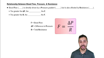
Relationship Between Blood Flow, Pressure, & Resistance