 Indietro
Chapter 1: A Preview of Cell Biology – Foundations, Methods, and Modern Approaches
Guida di studio - Note intelligenti
 Appunti personalizzati basati sui tuoi materiali, ampliati con definizioni chiave, esempi e contesto.
Introduction to Cell Biology
The Cell as the Fundamental Unit of Life
Cell biology is the study of cells, the basic structural and functional units of all living organisms. Modern cell biology integrates cytology, genetics, and biochemistry to understand the dynamic nature of cells.
Cells are the smallest units capable of independent life.
All living organisms are composed of one or more cells.
Cell biology is a rapidly advancing field due to technological and conceptual breakthroughs.
The Cell Theory: Historical Foundations
Development of the Cell Theory
The cell theory is a cornerstone of biology, describing the cellular basis of life. Its development was enabled by advances in microscopy and careful observation.
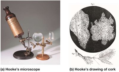
Robert Hooke (1665): First observed and named "cells" in cork tissue using a microscope.
Compound Microscopes (1830s): Improved magnification and resolution, allowing clearer observation of cell structures.
Key Contributors:
Robert Brown: Identified the nucleus in plant cells.
Matthias Schleiden: All plant tissues are composed of cells.
Thomas Schwann: All animal tissues are composed of cells.
Rudolf Virchow (1855): All cells arise from preexisting cells.
Three Tenets of Cell Theory:
All organisms consist of one or more cells.
The cell is the basic unit of structure for all organisms.
All cells arise only from preexisting cells.
The Emergence of Modern Cell Biology
Three Strands of Inquiry
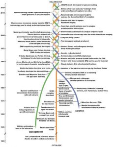
Modern cell biology is built upon three major disciplines:
Cytology: Focuses on cellular structure using optical techniques.
Biochemistry: Studies the chemistry of cellular structure and function.
Genetics: Investigates information flow, heredity, and genome sequencing.
Cytology: Cellular Structure and Microscopy
Microscopy and Cellular Dimensions
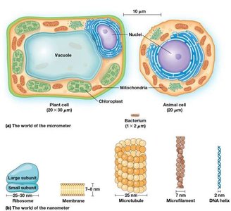
Microscopy is essential for studying cells due to their small size. Different units are used to measure cellular structures:
Micrometer (µm): 1 µm = 10−6 m; used for cells and organelles.
Nanometer (nm): 1 nm = 10−9 m; used for molecules and subcellular structures.
Angstrom (Å): 1 Å = 0.1 nm; used for atomic-scale measurements.
Types of Microscopy
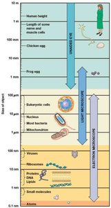
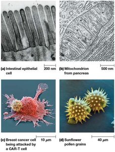
Light Microscopy (Brightfield): Uses visible light to observe stained, fixed cells. Resolution limit: 200–350 nm.
Specialized Light Microscopes: Phase-contrast, differential interference contrast, fluorescence, and confocal microscopy allow observation of living cells and specific molecules.
Electron Microscopy: Uses electron beams for much higher resolution (up to 100,000× magnification). Includes Transmission Electron Microscopy (TEM) and Scanning Electron Microscopy (SEM).
Biochemistry: Chemistry of Life
Biological Molecules and Pathways
Biochemistry explores the molecular basis of cellular structure and function.
Enzymes: Biological catalysts discovered through studies of fermentation (Pasteur, Buchners).
Metabolic Pathways: Glycolysis (Embden–Meyerhof pathway), Krebs cycle, and Calvin cycle elucidated in the early 20th century.
ATP: Identified as the principal energy storage molecule in cells.
Biochemical Methods
Subcellular Fractionation: Uses centrifugation to separate cell components.
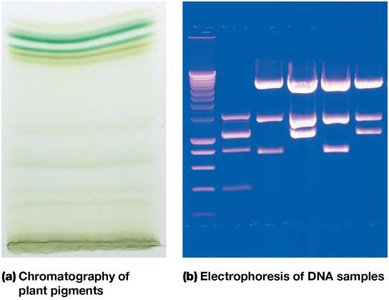
Chromatography: Separates molecules based on size, charge, or affinity.
Electrophoresis: Separates DNA, RNA, or proteins by size/charge in an electric field.
Mass Spectrometry: Determines the size and composition of proteins.
Genetics: Information Flow and Heredity
Classical and Molecular Genetics
Genetics investigates how traits are inherited and how genetic information is stored and expressed.
Mendelian Genetics: Gregor Mendel's pea experiments established the concept of genes as hereditary factors.
Chromosome Theory: Chromosomes carry genes; demonstrated by Morgan, Bridges, and Sturtevant using fruit flies (Drosophila melanogaster).
DNA as Genetic Material: DNA identified as the genetic material through experiments with bacteria and viruses (1940s).
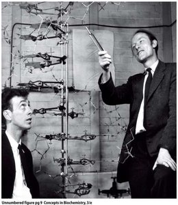
Watson and Crick (1953): Proposed the double helix model of DNA structure, aided by Rosalind Franklin's data.
Central Dogma of Molecular Biology
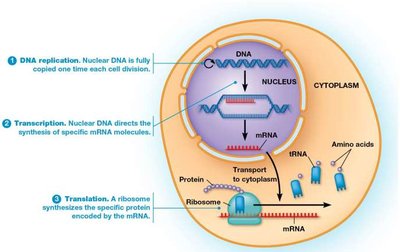
The central dogma describes the flow of genetic information in cells:
DNA → RNA → Protein
DNA is replicated, transcribed into RNA, and RNA is translated into protein.
Exceptions include some viruses and the process of reverse transcription.
Modern Genetic Techniques
Recombinant DNA Technology: Uses restriction enzymes to cut and recombine DNA from different sources.
DNA Cloning: Produces many copies of a specific DNA sequence.
DNA Sequencing: Determines the order of nucleotides in DNA; entire genomes can now be sequenced.
Bioinformatics: Uses computational tools to analyze large biological datasets (genomics, proteomics, transcriptomics, etc.).
CRISPR Genome Editing
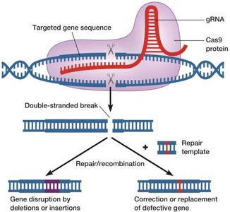
CRISPR is a revolutionary genome editing tool derived from a prokaryotic defense system.
Uses a guide RNA (gRNA) to target specific DNA sequences.
Cas9 protein introduces double-stranded breaks at the target site.
Repair can result in gene disruption or precise correction using a repair template (homology-directed repair).
Scientific Method and Model Organisms
Nature of Scientific Knowledge
Scientific facts are provisional and subject to change as new evidence emerges. Hypotheses are tested through controlled experiments, and models are developed to explain observations.
Hypothesis: A testable explanation for a phenomenon.
Null Hypothesis: The default assumption that there is no effect or relationship.
Controlled Experiments: Only one variable (independent variable) is changed at a time; the outcome is the dependent variable.
In vivo: Experiments in living organisms.
In vitro: Experiments outside living organisms (e.g., in test tubes).
Model Organisms in Cell Biology
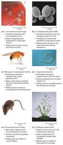
Model organisms are species that are widely studied, easy to manipulate, and provide insights applicable to other organisms.
Examples include Escherichia coli (bacterium), Saccharomyces cerevisiae (yeast), Drosophila melanogaster (fruit fly), Caenorhabditis elegans (nematode), Mus musculus (mouse), and Arabidopsis thaliana (plant).
Cell and tissue cultures are also used to study cellular processes in controlled environments.
Summary Table: Key Microscopy Techniques
Microscopy Type | Principle | Resolution | Application |
|---|---|---|---|
Light (Brightfield) | Visible light through stained specimen | 200–350 nm | General cell structure |
Phase-contrast/DIC | Enhances contrast in living cells | ~200 nm | Live cell imaging |
Fluorescence | Fluorescent tags/antibodies | ~200 nm | Specific molecules, proteins |
Confocal | Laser scans single plane | ~200 nm | 3D imaging, thick specimens |
Electron (TEM/SEM) | Electron beams | ~2 nm (TEM), ~10 nm (SEM) | Ultrastructure, surface details |
Additional info: This summary integrates foundational concepts from cytology, biochemistry, and genetics, as well as modern experimental approaches and the scientific method, providing a comprehensive overview suitable for introductory cell biology students.
