Ch. 8 - Molecular Biology of Transcription and RNA Processing
Sanders - Genetic Analysis: An Integrated Approach 3rd Edition
Sanders3rd EditionGenetic Analysis: An Integrated ApproachISBN: 9780135564172Non è quello che usi tu?Cambia libro di testo
Capitolo 8, Problema 19c

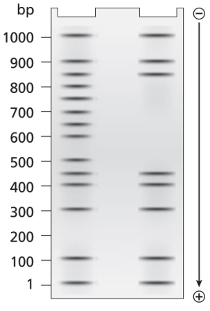
A 1.0-kb DNA fragment from the end of the mouse gene described in the previous problem is examined by DNA footprint protection analysis. Two samples are end-labeled with ³²P and one of the two is mixed with TFIIB, TFIID, and RNA polymerase II. The DNA exposed to these proteins is run in the right-hand lane of the gel shown below and the control DNA is run in the left-hand. Both DNA samples are treated with DNase I before running the samples on the electrophoresis gel.
Explain the role of DNase I.
Electrophoresis gel showing DNA fragments, with a ladder indicating sizes from 100 to 1000 base pairs.

Guida verificata passo dopo passo
1
DNase I is an enzyme that cleaves DNA by hydrolyzing the phosphodiester bonds between nucleotides. Its activity is nonspecific, meaning it cuts DNA at random locations unless the DNA is protected by bound proteins.
In DNA footprint protection analysis, DNase I is used to identify regions of DNA that are bound by proteins. These bound regions are protected from DNase I cleavage, creating a 'footprint' on the gel where no DNA fragments are observed.
The control DNA sample (left-hand lane) is treated with DNase I without any proteins present. This results in random cleavage across the entire DNA fragment, producing a ladder of DNA fragments of varying sizes.
The experimental DNA sample (right-hand lane) is treated with DNase I after being mixed with TFIIB, TFIID, and RNA polymerase II. These proteins bind to specific regions of the DNA, protecting those regions from DNase I cleavage.
By comparing the gel lanes, the protected regions (footprints) can be identified as areas where DNA fragments are missing in the experimental lane but present in the control lane. This reveals the binding sites of the proteins on the DNA.

Risposta video verificata per un problema simile:

Questa soluzione video è stata consigliata dai nostri tutor come utile per risolvere questo problema.
Durata del video:
2m

Concetti chiave

Ecco i concetti essenziali che devi comprendere per rispondere correttamente alla domanda.

DNase I Function

DNase I is an enzyme that cleaves the phosphodiester bonds between nucleotides in DNA, leading to the degradation of DNA into smaller fragments. In the context of DNA footprinting, DNase I is used to selectively digest unprotected regions of DNA, allowing researchers to identify which parts of the DNA are bound by proteins. This helps in understanding protein-DNA interactions and the regulatory mechanisms of gene expression.
Video consigliato:
Percorso guidato
08:26
Functional Genomics

DNA Footprinting

DNA footprinting is a technique used to study the binding of proteins to specific DNA sequences. By treating DNA with DNase I in the presence of bound proteins, researchers can create a 'footprint' that reveals protected regions of DNA where proteins are attached. The resulting pattern, observed through gel electrophoresis, indicates the locations of protein binding sites, providing insights into gene regulation and transcriptional control.
Video consigliato:
Percorso guidato
01:45
DNA Proofreading

Electrophoresis

Electrophoresis is a laboratory technique used to separate DNA fragments based on their size and charge. In this process, DNA samples are placed in a gel matrix and subjected to an electric field, causing the negatively charged DNA to migrate towards the positive electrode. The distance traveled by each fragment correlates with its size, allowing for the visualization and analysis of DNA fragments, including those generated by DNase I digestion in footprinting experiments.
Video consigliato:
Pratica correlata
Domanda del libro di testo

Wild-type E. coli grows best at 37°C but can grow efficiently up to 42°C. An E. coli strain has a mutation of the sigma subunit that results in an RNA polymerase holoenzyme that is stable and transcribes at wild-type levels at 37°C. The mutant holoenzyme is progressively destabilized as the temperature is raised, and it completely denatures and ceases to carry out transcription at 42°C. Relative to wild-type growth, characterize the ability of the mutant strain to carry out transcription at 40°C

867
views
Domanda del libro di testo

Wild-type E. coli grow best at 37°C but can grow efficiently up to 42°C. An E. coli strain has a mutation of the sigma subunit that results in an RNA polymerase holoenzyme that is stable and transcribes at wild-type levels at 37°C. The mutant holoenzyme is progressively destabilized as the temperature is raised, and it completely denatures and ceases to carry out transcription at 42°C. Relative to wild-type growth, characterize the ability of the mutant strain to carry out transcription at 37°C

482
views
Domanda del libro di testo

Wild-type E. coli grows best at 37°C but can grow efficiently up to 42°C. An E. coli strain has a mutation of the sigma subunit that results in an RNA polymerase holoenzyme that is stable and transcribes at wild-type levels at 37°C. The mutant holoenzyme is progressively destabilized as the temperature is raised, and it completely denatures and ceases to carry out transcription at 42°C. Relative to wild-type growth, characterize the ability of the mutant strain to carry out transcription at 42°C

554
views
Domanda del libro di testo

A 3.5-kb segment of DNA containing the complete sequence of a mouse gene is available. The DNA segment contains the promoter sequence and extends beyond the polyadenylation site of the gene. The DNA is studied by band shift assay, and the following gel bands are observed.

Match these conditions to a specific lane of the gel.

3.5-kb fragment plus TFIIB

473
views
Domanda del libro di testo

A 1.0-kb DNA fragment from the end of the mouse gene described in the previous problem is examined by DNA footprint protection analysis. Two samples are end-labeled with ³²P and one of the two is mixed with TFIIB, TFIID, and RNA polymerase II. The DNA exposed to these proteins is run in the right-hand lane of the gel shown below and the control DNA is run in the left-hand. Both DNA samples are treated with DNase I before running the samples on the electrophoresis gel.

What length of DNA is bound by the transcriptional proteins? Explain how the gel results support this interpretation.

561
views
Domanda del libro di testo

A 1.0-kb DNA fragment from the end of the mouse gene described in the previous problem is examined by DNA footprint protection analysis. Two samples are end-labeled with ³²P, and one of the two is mixed with TFIIB, TFIID, and RNA polymerase II. The DNA exposed to these proteins is run in the right-hand lane of the gel shown below and the control DNA is run in the left-hand. Both DNA samples are treated with DNase I before running the samples on the electrophoresis gel.

Draw a diagram of this DNA fragment bound by the transcriptional proteins, showing the approximate position of proteins along the fragment.

472
views