 뒤로
Autonomic Nervous System: Structure, Function, and Clinical Relevance
스터디 가이드 - 스마트 노트
 자료에 맞춘 맞춤형 노트, 핵심 정의, 예시, 맥락을 확장해 제공합니다.
Autonomic Nervous System (ANS)
Introduction to the ANS
The Autonomic Nervous System (ANS) is a division of the peripheral nervous system responsible for involuntary physiological functions. It operates outside conscious control, regulating processes such as body temperature, cardiovascular, respiratory, digestive, excretory, and reproductive functions. The ANS enables survival in coma patients by maintaining essential bodily functions.
Key Functions: Routine adjustment of internal systems, maintaining homeostasis.
Effectors: Smooth muscle, cardiac muscle, glands, and adipose tissue.
Clinical Relevance: Dysfunction can lead to disorders such as Raynaud’s Disease.
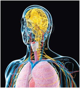
Functional Overview of the Nervous System
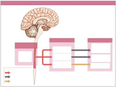
The nervous system is divided into the Central Nervous System (CNS) and Peripheral Nervous System (PNS). The PNS includes the Somatic Nervous System (SNS) and the Autonomic Nervous System (ANS). The ANS itself is subdivided into the Sympathetic and Parasympathetic divisions, which often have opposing effects on target organs.
SNS: Controls voluntary movements via skeletal muscles.
ANS: Controls involuntary functions via visceral organs.
Motor Pathways: ANS uses two-neuron chains: preganglionic (myelinated) and postganglionic (unmyelinated).
Sympathetic Division (Thoracolumbar Division)
Organization and Pathways
The Sympathetic Division prepares the body for 'fight-or-flight' responses during stress or emergency. Preganglionic neurons originate from spinal segments T1–L2, with cell bodies in the lateral gray horns. Their axons pass through ventral roots to ganglia near the vertebral column.
Sympathetic Chain Ganglia: Paired structures alongside the vertebral column, innervating body wall, limbs, and thoracic organs.
Collateral Ganglia: Unpaired structures anterior to the aorta, innervating abdominopelvic organs.
Adrenal Medulla: Specialized neurons release hormones (epinephrine and norepinephrine) into the bloodstream.
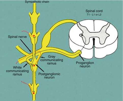
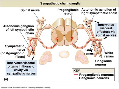
Sympathetic Chain Ganglia: Pathways
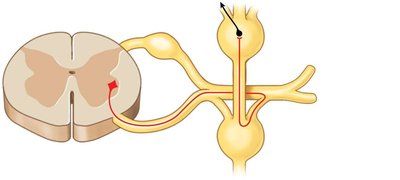
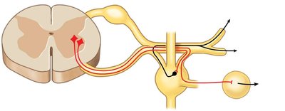
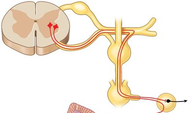
Preganglionic fibers enter the sympathetic chain ganglia via the white ramus communicans (myelinated). Postganglionic fibers exit via the gray ramus communicans (unmyelinated). Fibers may synapse at the same level, ascend or descend before synapsing, or pass through to collateral ganglia.
Option 1A: Postganglionic fibers re-enter spinal nerve to innervate body wall and limbs.
Option 1B: Postganglionic fibers exit as sympathetic nerves to innervate thoracic organs.
Option 2: Preganglionic fibers pass through chain ganglia to collateral ganglia for abdominopelvic organs.
Collateral Ganglia
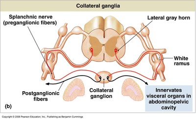
Collateral ganglia are sites where preganglionic neurons innervating abdominopelvic viscera synapse. Postganglionic fibers form splanchnic nerves, which are variable in appearance and location.
Celiac Ganglion: Greater splanchnic nerve; innervates digestive system and kidney.
Superior Mesenteric Ganglion: Lesser splanchnic nerve; innervates small and large intestine.
Inferior Mesenteric Ganglion: Lumbar splanchnic nerve; innervates large intestine, kidney, bladder, and sex organs.
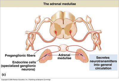
Adrenal Medulla
Some preganglionic fibers bypass the sympathetic chain and synapse on modified neurons in the adrenal medulla. These cells release epinephrine (E) and norepinephrine (NE) directly into the bloodstream, acting as hormones to amplify sympathetic effects throughout the body.
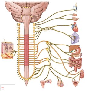
Effects of Sympathetic Stimulation
Increased alertness
Enhanced cardiovascular and respiratory activity
Increased muscle tone
Mobilization of energy reserves
Redistribution of blood to skeletal muscle
Parasympathetic Division (Craniosacral Division)
Organization and Pathways
The Parasympathetic Division promotes 'rest-and-digest' activities, supporting relaxation, food processing, and energy absorption. Preganglionic neurons originate in the brainstem (cranial nerves III, VII, IX, X) and sacral spinal segments (S2–S4). Ganglionic neurons are located close to or within target organs.
Cranial Nerves: N III, VII, IX innervate head structures; N X (Vagus) innervates thoracic and abdominopelvic organs.
Sacral Outflow: S2–S4 form pelvic nerves, innervating lower abdominopelvic organs.
Effects: More specific and localized than sympathetic division.
Major Effects of Parasympathetic Stimulation
Pupil constriction
Secretion of digestive enzymes and hormones
Increased smooth muscle activity in digestive tract
Stimulation of defecation and urination
Constriction of respiratory passages
Reduced heart rate and force of contraction
Sexual arousal
Synapses and Neurotransmitter Release
Sympathetic Division
Preganglionic fibers: Short, myelinated; release acetylcholine (ACh).
Postganglionic fibers: Long, unmyelinated; release norepinephrine (NE) (except ACh for sweat glands).
Adrenal gland: Releases epinephrine (E) and norepinephrine (NE) as hormones.
Parasympathetic Division
Preganglionic fibers: Long, myelinated; release acetylcholine (ACh).
Postganglionic fibers: Short, unmyelinated; release acetylcholine (ACh).
Comparison of Sympathetic and Parasympathetic Divisions
Feature | Sympathetic | Parasympathetic |
|---|---|---|
Origin | T1–L2 (thoracolumbar) | Brainstem, S2–S4 (craniosacral) |
Preganglionic Fiber | Short, myelinated | Long, myelinated |
Postganglionic Fiber | Long, unmyelinated | Short, unmyelinated |
Neurotransmitter | ACh (preganglionic), NE (postganglionic) | ACh (both) |
General Effect | Fight-or-flight | Rest-and-digest |
Clinical Anatomy: Raynaud’s Disease
Raynaud’s Disease
Raynaud’s Disease is a clinical disorder involving excessive sympathetic constriction of small arteries in the fingers and toes, typically in response to cold or stress. This results in reduced blood flow, causing the affected areas to turn white or blue, loss of sensation, pain, and increased risk of frostbite.
Symptoms: Color changes, numbness, pain.
Risk: Increased susceptibility to tissue damage in cold environments.
Key Terms and Definitions
Preganglionic Neuron: The first neuron in the two-neuron chain, originating in the CNS.
Postganglionic Neuron: The second neuron, located in a ganglion, projecting to the effector organ.
Ganglion: A cluster of neuronal cell bodies in the PNS.
White Ramus Communicans: Myelinated pathway for preganglionic fibers entering the sympathetic chain.
Gray Ramus Communicans: Unmyelinated pathway for postganglionic fibers exiting the sympathetic chain.
Splanchnic Nerve: Nerve containing autonomic fibers, often associated with collateral ganglia.
Adrenal Medulla: Endocrine organ releasing hormones in response to sympathetic stimulation.
Additional info:
The Enteric Nervous System (ENS) is sometimes considered a third division of the ANS, controlling gastrointestinal function independently.
Sympathetic and parasympathetic divisions can act independently or together, depending on the organ system and physiological context.
