 뒤로
BIO117 Unit 3 Study Guide – Anatomy & Physiology Guidance
스터디 가이드 - 스마트 노트
 자료에 맞춘 맞춤형 노트, 핵심 정의, 예시, 맥락을 확장해 제공합니다.
Q1. Match the 3 Types of Neurons to their descriptions: Sensory neurons, Interneurons, Motor neurons
Background
Topic: Nervous System – Neuron Types
This question tests your understanding of the functional roles of different neuron types in the nervous system.
Key Terms:
Sensory neurons: Carry information from sensory receptors to the central nervous system.
Interneurons: Process information within the central nervous system; connect sensory and motor neurons.
Motor neurons: Transmit signals from the central nervous system to effectors (muscles or glands).
Step-by-Step Guidance
Review the definitions and functions of each neuron type.
Think about the direction of information flow: sensory neurons bring information in, interneurons process it, motor neurons send instructions out.
Match each neuron type to its description based on its role in the nervous system.
Try solving on your own before revealing the answer!
Final Answer:
Sensory neurons: Carry information from receptors to CNS
Interneurons: Connect sensory and motor neurons within CNS
Motor neurons: Carry instructions from CNS to effectors
Each neuron type has a distinct role in the nervous system's communication pathways.
Q2. Match the Cerebral Hemispheres with their functions: Frontal lobe, Parietal lobe, Occipital lobe, Temporal lobe
Background
Topic: Brain Anatomy – Cerebral Lobes
This question tests your knowledge of the functional specialization of the brain's lobes.
Key Terms:
Frontal lobe: Associated with reasoning, planning, movement, and problem-solving.
Parietal lobe: Processes sensory information such as touch, temperature, and pain.
Occipital lobe: Responsible for visual processing.
Temporal lobe: Involved in hearing, memory, and language.
Step-by-Step Guidance
Recall the main functions associated with each lobe.
Match each lobe to its primary function based on its anatomical location and role.
Consider examples: vision (occipital), touch (parietal), movement (frontal), hearing (temporal).
Try solving on your own before revealing the answer!
Final Answer:
Frontal lobe: Reasoning, movement, problem-solving
Parietal lobe: Sensory processing
Occipital lobe: Visual processing
Temporal lobe: Hearing, memory, language
Each lobe is specialized for distinct cognitive and sensory functions.
Q3. Match the part of Diencephalon with its function: Hypothalamus, Thalamus, Pineal gland, Pituitary gland
Background
Topic: Brain Anatomy – Diencephalon
This question tests your understanding of the functions of key diencephalon structures.
Key Terms:
Hypothalamus: Regulates homeostasis, controls pituitary gland.
Thalamus: Relay station for sensory information.
Pineal gland: Produces melatonin, regulates sleep cycles.
Pituitary gland: Secretes hormones that regulate other glands.
Step-by-Step Guidance
Review the main function of each structure.
Match each part to its description based on its role in endocrine and nervous system regulation.
Think about which structure is involved in hormone production, sensory relay, or homeostasis.
Try solving on your own before revealing the answer!
Final Answer:
Hypothalamus: Regulates homeostasis, controls pituitary
Thalamus: Sensory relay station
Pineal gland: Produces melatonin
Pituitary gland: Secretes hormones
The diencephalon integrates sensory, endocrine, and homeostatic functions.
Q4. Put the Reflex Arc pathway in order
Background
Topic: Nervous System – Reflex Arc
This question tests your understanding of the sequence of events in a reflex response.
Key Terms:
Reflex arc: The neural pathway involved in a reflex action.
Effector: Muscle or gland that responds to the motor neuron signal.
Step-by-Step Guidance
Identify the starting point: stimulus received by a receptor.
Follow the signal as it travels through the sensory neuron to the CNS.
Recognize the role of the interneuron in transmitting the signal to the motor neuron.
Consider the final step: effector responds to the motor neuron signal.
Try solving on your own before revealing the answer!
Final Answer:
Stimulus is received from a receptor
Sensory neuron transmits signal from receptor
An interneuron transmits signal directly to motor neuron
Motor neuron receives signal from interneuron
Effector responds
The reflex arc is a rapid, automatic response pathway.
Q5. Put the steps of Synaptic Transmission in order
Background
Topic: Nervous System – Synaptic Transmission
This question tests your understanding of the sequence of events during neurotransmitter release and signal transmission between neurons.
Key Terms:
Synaptic vesicles: Store neurotransmitters in the presynaptic neuron.
Synaptic cleft: The gap between neurons where neurotransmitters are released.
Postsynaptic neuron: Receives the neurotransmitter signal.
Step-by-Step Guidance
Start with the neuronal impulse traveling down the presynaptic axon.
Synaptic vesicles fuse with the axon bulb membrane.
Neurotransmitter is released into the synaptic cleft.
Neurotransmitters bind to receptors on the postsynaptic neuron.
Sodium ion channels open, allowing ions to enter the postsynaptic neuron.
Try solving on your own before revealing the answer!
Final Answer:
Neuronal impulse travels down the presynaptic axon to the axon bulb
Synaptic vesicles fuse with the axon bulb plasma membrane
Neurotransmitter is released into the synaptic cleft
Neurotransmitters bind to receptors on the postsynaptic neuron
Sodium ion channels open and the ions enter the postsynaptic neuron
This sequence enables communication between neurons.
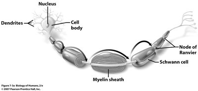
Q6. Label the parts of a Neuron: Axon bulb, Axon, Body, Dendrites, Myelin sheath, Schwann cell, Node of Ranvier
Background
Topic: Nervous System – Neuron Structure
This question tests your ability to identify and label the structural components of a neuron.
Key Terms:
Dendrites: Receive signals from other neurons.
Cell body: Contains the nucleus and organelles.
Axon: Transmits electrical impulses away from the cell body.
Myelin sheath: Insulates the axon, speeds up transmission.
Schwann cell: Produces myelin in the peripheral nervous system.
Node of Ranvier: Gaps in the myelin sheath where ion exchange occurs.
Axon bulb: End of the axon, releases neurotransmitters.
Step-by-Step Guidance
Examine the diagram and locate each labeled part.
Identify the dendrites, cell body, and axon based on their position and function.
Find the myelin sheath, Schwann cell, and Node of Ranvier along the axon.
Locate the axon bulb at the end of the axon.
Try labeling on your own before revealing the answer!
Final Answer:
Dendrites: Branches receiving signals
Cell body: Central area with nucleus
Axon: Long fiber transmitting impulses
Myelin sheath: Insulating layer
Schwann cell: Myelin-producing cell
Node of Ranvier: Gap in myelin sheath
Axon bulb: End of axon
Each part plays a specific role in neuron function and signal transmission.
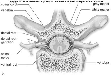
Q7. Label the parts of the Spinal column: Central canal, Dorsal root, Grey matter, Spinal nerve, Ventral root, Vertebrae, White matter
Background
Topic: Nervous System – Spinal Cord Anatomy
This question tests your ability to identify and label the structural components of the spinal cord and surrounding vertebrae.
Key Terms:
Central canal: Contains cerebrospinal fluid.
Dorsal root: Sensory input to spinal cord.
Ventral root: Motor output from spinal cord.
Grey matter: Contains neuron cell bodies.
White matter: Contains myelinated axons.
Spinal nerve: Mixed nerve carrying both sensory and motor information.
Vertebrae: Bones protecting the spinal cord.
Step-by-Step Guidance
Examine the diagram and locate each labeled part.
Identify the central canal, grey matter, and white matter within the spinal cord.
Find the dorsal root, ventral root, and spinal nerve based on their position and function.
Locate the vertebrae surrounding the spinal cord.
Try labeling on your own before revealing the answer!
Final Answer:
Central canal: Middle channel with CSF
Dorsal root: Sensory input
Ventral root: Motor output
Grey matter: Inner butterfly-shaped area
White matter: Outer area
Spinal nerve: Mixed nerve
Vertebrae: Surrounding bone
Each part is essential for spinal cord function and protection.
Q8. Match the endocrine glands with their products: Pineal, Thymus, Thyroid, Anterior Pituitary, Posterior Pituitary, Adrenal Cortex, Adrenal Medulla
Background
Topic: Endocrine System – Glands and Hormones
This question tests your knowledge of which hormones are produced by specific endocrine glands.
Key Terms:
Pineal: Melatonin
Thymus: Thymosins
Thyroid: Thyroxin
Anterior Pituitary: Growth Hormone
Posterior Pituitary: Antidiuretic Hormone
Adrenal Cortex: Mineralocorticoids
Adrenal Medulla: Norepinephrine
Step-by-Step Guidance
Review the hormones produced by each gland.
Match each gland to its primary hormone product.
Consider the function of each hormone in the body.
Try matching on your own before revealing the answer!
Final Answer:
Pineal: Melatonin
Thymus: Thymosins
Thyroid: Thyroxin
Anterior Pituitary: Growth Hormone
Posterior Pituitary: Antidiuretic Hormone
Adrenal Cortex: Mineralocorticoids
Adrenal Medulla: Norepinephrine
Each gland produces hormones essential for body regulation.
Q9. Glucose metabolism, with word bank: Adipose tissue, Glycogen, Glucagon, Insulin, Liver, Muscle cells, Pancreas
Background
Topic: Endocrine System – Glucose Metabolism
This question tests your understanding of how glucose is regulated in the body and the roles of various tissues and hormones.
Key Terms:
Insulin: Lowers blood glucose by promoting uptake into cells.
Glucagon: Raises blood glucose by promoting glycogen breakdown.
Liver: Stores and releases glucose.
Muscle cells: Store glucose as glycogen.
Adipose tissue: Stores excess glucose as fat.
Pancreas: Produces insulin and glucagon.
Glycogen: Storage form of glucose.
Step-by-Step Guidance
Identify the role of insulin and glucagon in glucose regulation.
Consider how the liver, muscle cells, and adipose tissue participate in glucose storage and release.
Use the word bank to fill in the blanks for each step in glucose metabolism.
Try completing the steps before revealing the answer!
Final Answer:
Insulin: Promotes glucose uptake
Glucagon: Promotes glycogen breakdown
Liver: Stores/releases glucose
Muscle cells: Store glycogen
Adipose tissue: Stores fat
Pancreas: Produces hormones
Glycogen: Storage form
Glucose metabolism involves coordinated actions of hormones and tissues.
Q10. Calcium metabolism, with word bank: Calcitonin, Homeostasis, Osteoclasts, Osteoblasts, Parathyroid, Parathyroid hormone, Thyroid
Background
Topic: Endocrine System – Calcium Regulation
This question tests your understanding of how calcium levels are regulated in the body and the roles of hormones and cells.
Key Terms:
Calcitonin: Lowers blood calcium.
Parathyroid hormone: Raises blood calcium.
Osteoclasts: Break down bone, release calcium.
Osteoblasts: Build bone, use calcium.
Thyroid: Produces calcitonin.
Parathyroid: Produces parathyroid hormone.
Homeostasis: Maintenance of stable internal environment.
Step-by-Step Guidance
Identify the hormones involved in calcium regulation.
Consider the roles of osteoclasts and osteoblasts in bone remodeling.
Use the word bank to fill in the blanks for each step in calcium metabolism.
Try completing the steps before revealing the answer!
Final Answer:
Calcitonin: Lowers calcium
Parathyroid hormone: Raises calcium
Osteoclasts: Release calcium
Osteoblasts: Use calcium
Thyroid: Calcitonin source
Parathyroid: Parathyroid hormone source
Homeostasis: Stable calcium levels
Calcium metabolism is regulated by hormones and bone cells to maintain homeostasis.
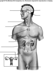
Q11. Label the locations of the glands on the body: Adrenal gland, Gonads, Hypothalamus, Pancreas, Pineal Gland, Pituitary Gland, Thymus, Thyroid
Background
Topic: Endocrine System – Gland Locations
This question tests your ability to identify the anatomical locations of major endocrine glands.
Key Terms:
Adrenal gland: Above kidneys
Gonads: Ovaries/testes
Hypothalamus: Base of brain
Pancreas: Behind stomach
Pineal gland: Deep in brain
Pituitary gland: Below hypothalamus
Thymus: Upper chest
Thyroid: Neck
Step-by-Step Guidance
Examine the diagram and locate each gland based on anatomical landmarks.
Identify the glands in the brain, neck, chest, abdomen, and pelvis.
Label each gland according to its position in the body.
Try labeling on your own before revealing the answer!
Final Answer:
Adrenal gland: Above kidneys
Gonads: Ovaries/testes
Hypothalamus: Base of brain
Pancreas: Behind stomach
Pineal Gland: Deep in brain
Pituitary Gland: Below hypothalamus
Thymus: Upper chest
Thyroid: Neck
Each gland is located in a specific region, essential for hormone production and regulation.
