 뒤로
Blood and Blood Vessels: Structure, Function, and Physiology
스터디 가이드 - 스마트 노트
 자료에 맞춘 맞춤형 노트, 핵심 정의, 예시, 맥락을 확장해 제공합니다.
Cardiovascular System Overview and Blood Properties
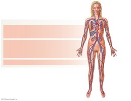
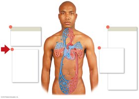
Components of the Cardiovascular System
The cardiovascular system is responsible for the transport of blood throughout the body, ensuring the delivery of oxygen, nutrients, and the removal of waste products. It consists of three main components: blood, blood vessels, and the heart.
Blood: The fluid medium for transport.
Blood Vessels: Arteries, veins, and capillaries distribute blood.
Heart: The muscular pump that propels blood and maintains pressure.
Functions of Blood
Blood is a fluid connective tissue with several essential functions:
Transportation: Moves respiratory gases, nutrients, waste, and hormones.
Regulation: Maintains pH and ion concentration.
Restriction of Fluid Loss: Clotting mechanisms prevent excessive bleeding.
Defense: Contains white blood cells and antibodies for immunity.
Thermoregulation: Distributes heat due to its high heat capacity.
Components of Blood
Blood Composition
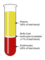
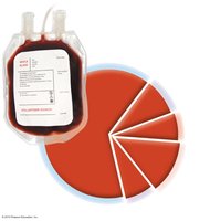
Blood consists of plasma and formed elements. Plasma is the liquid portion, while formed elements include cells and cell fragments.
Plasma: 55% of total blood volume; mostly water, with proteins and solutes.
Buffy Coat: Less than 1%; contains leukocytes (white blood cells) and platelets.
Erythrocytes: 45% of total blood volume; red blood cells.
Plasma
Plasma is the fluid component of blood, composed of:
Water: 92% of plasma.
Plasma Proteins: 7% (albumins, globulins, fibrinogen).
Other Solutes: 1% (electrolytes, nutrients, wastes).
Formed Elements
Formed elements include erythrocytes, leukocytes, and platelets.
Erythrocytes (Red Blood Cells): Responsible for oxygen transport.
Leukocytes (White Blood Cells): Immune functions.
Platelets: Essential for blood clotting.
Red Blood Cells (Erythrocytes)
Structure and Function
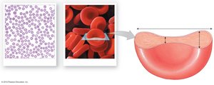
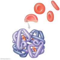
Red blood cells are small, biconcave discs that maximize surface area for gas exchange. They lack nuclei and organelles, containing only hemoglobin for oxygen transport.
Hematocrit: Percentage of blood composed of RBCs (typically ~45%).
Lifespan: Approximately 120 days.
Major Purpose: Oxygen transport.
Hemoglobin
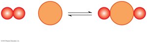
Hemoglobin is the protein within RBCs that binds oxygen. Each molecule consists of four globin subunits, each with an iron-containing heme group.
Oxyhemoglobin (HbO2): Formed when O2 binds to heme; bright red color.
Deoxyhemoglobin: Dark red color.
White Blood Cells (Leukocytes)
Types and Functions
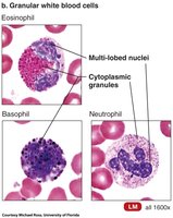
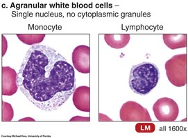
White blood cells are crucial for immune defense. They are classified as granular or agranular based on the presence of cytoplasmic granules.
Granular Leukocytes: Neutrophils, eosinophils, basophils.
Agranular Leukocytes: Monocytes, lymphocytes.
Movement and Response
Amoeboid Motion: Self-propelled movement.
Diapedesis: Squeezing through vessel walls.
Positive Chemotaxis: Movement toward chemical signals from damaged tissue.
Platelets
Structure and Function
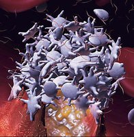
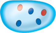
Platelets are cell fragments derived from megakaryocytes. They play a vital role in hemostasis and clot formation.
Lifespan: 9-12 days.
Function: Initiate clotting and repair damaged vessels.
Hematopoiesis and Hemostasis
Hematopoiesis
Hematopoiesis is the process of forming new blood cells in red bone marrow. Hemocytoblasts are multipotent stem cells that differentiate into various blood cell types.
Lymphoid Stem Cells: Produce lymphocytes.
Myeloid Stem Cells: Produce all other formed elements.
Regulation: Colony stimulating factors and erythropoietin (EPO) control production.
Hemostasis
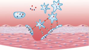
Hemostasis is the process of stopping bleeding, involving three phases:
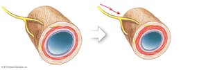
Vascular Phase: Vessel constriction and exposure of basement membrane.
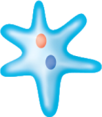
Platelet Phase: Platelets adhere to damaged area and release chemicals.
Coagulation Phase: Formation of a fibrin mesh to stabilize the clot.
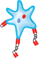
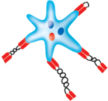
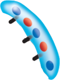
Coagulation Phase
The coagulation phase involves a cascade of clotting factors, leading to the activation of Factor X and the formation of prothrombinase. Prothrombinase converts prothrombin to thrombin, which then converts fibrinogen to fibrin.
Extrinsic Pathway: Triggered by external tissue damage.
Intrinsic Pathway: Triggered by damage within the vessel.
Common Pathway: Both pathways converge to activate Factor X.
Clot Retraction and Fibrinolysis
Clot retraction pulls wound edges together. Fibrinolysis, mediated by plasmin, breaks down the fibrin mesh after healing.
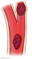
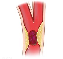
Embolus: Drifting blood clot.
Embolism: Blockage caused by an embolus.
Thrombus: Clot attached to vessel wall.
Blood Types
Antigens and Antibodies
Blood types are determined by antigens on the surface of red blood cells. The ABO group and Rh factor are the most clinically significant.
Type A: Surface antigen A; anti-B antibodies.
Type B: Surface antigen B; anti-A antibodies.
Type AB: Both antigens; no anti-A or anti-B antibodies.
Type O: No antigens; both anti-A and anti-B antibodies.
Agglutination and Hemolysis
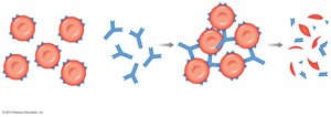
When opposing antibodies encounter their antigen, agglutination (clumping) occurs, which can lead to hemolysis and dangerous complications.
Rh Factor and Hemolytic Disease of the Newborn
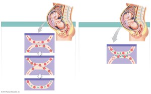
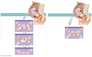
Rh-negative individuals can develop anti-Rh antibodies after exposure to Rh-positive blood. In pregnancy, this can cause erythroblastosis fetalis if the mother is Rh-negative and the fetus is Rh-positive.
Blood Vessel Structure
Types of Blood Vessels
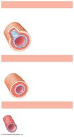
Blood vessels are classified as arteries, veins, and capillaries. Each type has distinct structural features.
Arteries: Carry blood away from the heart; thick walls, high pressure.
Veins: Return blood to the heart; thinner walls, lower pressure, contain valves.
Capillaries: Sites of exchange; extremely thin walls.
Blood Vessel Layers
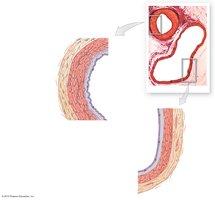
Blood vessels have three main layers:
Tunica intima: Innermost layer; endothelium and connective tissue.
Tunica media: Middle layer; smooth muscle and elastic fibers.
Tunica externa: Outer layer; connective tissue sheath.
Comparison of Arteries and Veins
Arteries and veins differ in structure and function. The following table summarizes their features:
Feature | Typical Artery | Typical Vein |
|---|---|---|
General appearance in sectional view | Usually round, thick wall | Usually flattened or collapsed, thin wall |
Tunica intima | Usually rippled | Often smooth |
Internal elastic membrane | Present | Absent |
Tunica media | Thick, dominated by smooth muscle | Thin, dominated by smooth muscle and collagen |
External elastic membrane | Present | Absent |
Tunica externa | Collagen and elastic fibers | Collagen and elastic fibers, smooth muscle |
Capillaries and Capillary Beds
Structure and Function
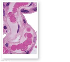
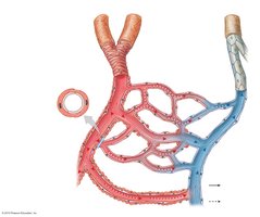
Capillaries are the only vessels where exchange of gases, nutrients, and wastes occurs. They may have fenestrae (pores) to facilitate diffusion.
Capillary Beds and Regulation
Capillaries form networks called beds, with flow regulated by precapillary sphincters. Anastomoses allow blood to bypass capillary beds when needed.
Circulation Patterns
Pulmonary and Systemic Circuits
The cardiovascular system is divided into pulmonary and systemic circuits:
Pulmonary Circuit: Carries blood to the lungs for oxygenation.
Systemic Circuit: Delivers oxygenated blood to body tissues.
Venous Return and Valves
Veins contain valves to prevent backflow, especially since blood pressure is lower. Skeletal muscle contractions help move blood toward the heart.
Summary
The blood and blood vessels are integral to the cardiovascular system, facilitating transport, regulation, defense, and homeostasis. Understanding their structure and function is essential for comprehending human physiology.
