 뒤로
L20 Blood Flow
스터디 가이드 - 스마트 노트
 자료에 맞춘 맞춤형 노트, 핵심 정의, 예시, 맥락을 확장해 제공합니다.
Blood Flow and Cardiac Output
Stroke Volume and Afterload
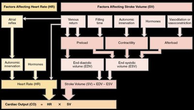
Stroke volume (SV) is the amount of blood ejected by the ventricle in one contraction. It is determined by the difference between end-diastolic volume (EDV) and end-systolic volume (ESV):
Stroke Volume (SV):
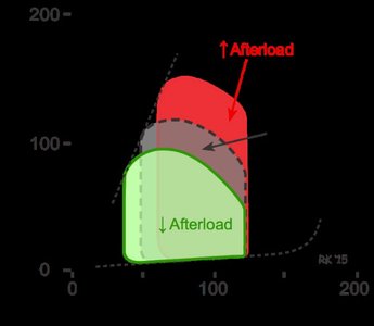
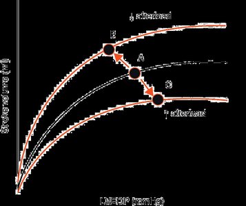
Afterload: The resistance the heart must overcome to eject blood. Increased afterload reduces stroke volume, while afterload reduction increases SV without increasing oxygen requirement.
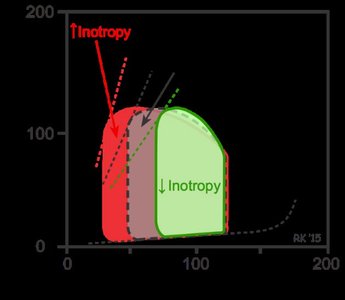
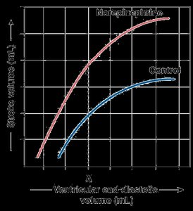
Contractility: The intrinsic ability of cardiac muscle to contract. Influenced by catecholamines (e.g., norepinephrine), increased Ca2+ permeability, and muscle stretch (increased filling).
Cardiac Output and Regulation
Cardiac output (CO) is the volume of blood pumped by the heart per minute. It is calculated as:
Cardiac Output (CO):
Normal Output: Approximately 4.9 L/min in adults.
Autonomic Regulation:
Sympathetic stimulation increases heart rate (HR) via β-adrenergic receptors and catecholamines.
Parasympathetic stimulation decreases HR via muscarinic acetylcholine receptors (mAChR).
Atropine is an mAChR antagonist, blocking parasympathetic effects.
Factors Affecting Cardiac Output
Cardiac output is influenced by heart rate and stroke volume, which are regulated by multiple factors:
Heart Rate: Controlled by autonomic innervation and hormones.
Stroke Volume: Influenced by preload (venous return), contractility, and afterload.
Preload: Determined by venous return, filling time, skeletal muscle pump, respiratory pump, venoconstriction, atrial systole, venous pressure, and cardiac suction.
Afterload: Determined by diastolic blood pressure (DBP).
Contractility: Increased by sympathetic stimulation and Ca2+ permeability.
Blood Pressure Determinants
Blood pressure is determined by cardiac performance and vascular resistance:
Systolic Blood Pressure (SBP): Chiefly determined by stroke volume and cardiac performance.
Diastolic Blood Pressure (DBP): Chiefly determined by vascular resistance and flow during diastole.
Mean Arterial Pressure (MAP): or
Pulse Pressure (PP):
Ejection Fraction (EF):
Baroreceptor Reflex
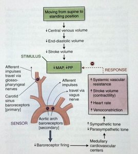
The baroreceptor reflex helps maintain blood pressure by adjusting heart rate, stroke volume, and vascular resistance in response to changes in blood pressure.
Capillaries and Capillary Exchange
Structure and Function of Capillaries
Capillaries are the smallest blood vessels, consisting of a single layer of endothelial cells. They facilitate exchange of nutrients, gases, and waste between blood and tissues.
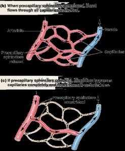
Precapillary Sphincters: Smooth muscle structures that regulate blood flow into capillary beds in response to local signals.
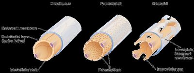
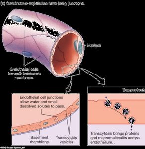
Endothelial Cells: Form the capillary wall; types include continuous, fenestrated, and sinusoidal.
Pericytes: Support capillary structure, decrease permeability, and can differentiate into other cell types.
Metarteriole: Provides a bypass route for blood flow.
Capillary Exchange Mechanisms
Three primary mechanisms facilitate exchange between interstitial fluid (ISF) and plasma:
Diffusion: Movement of molecules from high to low concentration; main method for nutrients and gases.
Vesicle Transport: Substances move across cell membranes in vesicles via endocytosis and exocytosis.
Bulk Flow: Movement of protein-free plasma through water-filled channels; distributes extracellular fluid between plasma and ISF.
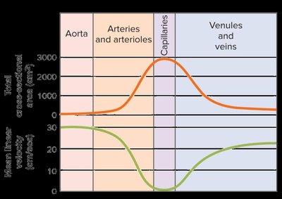
Capillary Exchange: Pressure Differences
Exchange across capillary walls is driven by hydrostatic and osmotic pressures:
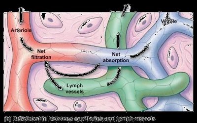
Arterial End: Filtration occurs due to higher hydrostatic pressure (net positive pressure).
Venule End: Absorption occurs due to higher colloid osmotic pressure (net negative pressure).
Lymphatic System: Absorbs excess fluid, preventing edema.
Edema: Results from increased hydrostatic pressure or impaired lymphatic drainage.
Lymphatic System
Structure and Function
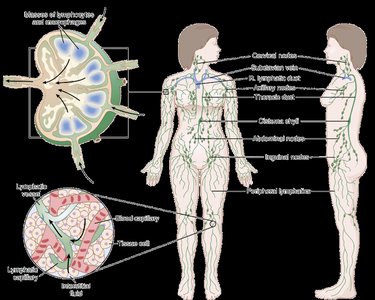
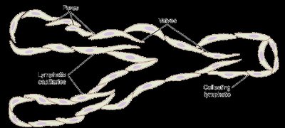
The lymphatic system is an accessory route for excess interstitial fluid and proteins, playing a crucial role in preventing edema and supporting immune function.
Functions:
Prevents edema by returning excess ISF to the bloodstream.
Supports immune system by transporting lymphocytes and antigen-presenting cells.
Absorbs nutrients from the gastrointestinal tract.
Lymphatic Pump:
Contraction of smooth muscle filaments in lymph vessels propels lymph.
External compression (e.g., skeletal muscle activity) enhances lymphatic pumping.
Key Equations and Relationships
(flow)
Glossary of Key Terms
HR: Heart rate
SNS: Sympathetic nervous system
PSNS: Parasympathetic nervous system
TPR: Total peripheral resistance
ΔP: Change in pressure
Q or F: Flow
R: Resistance
l: Length
η: Viscosity
r: Radius
v: Velocity
CSA: Cross-sectional area
Ang II: Angiotensin II
MAP: Mean arterial pressure
CO: Cardiac output
DBP: Diastolic blood pressure
SBP: Systolic blood pressure
PP: Pulse pressure
EF: Ejection fraction
SV: Stroke volume
EDV: End diastolic volume
ESV: End systolic volume
