 뒤로
뒤로Blood: Structure, Function, and Disorders – Study Notes for Anatomy & Physiology
스터디 가이드 - 스마트 노트
 자료에 맞춘 맞춤형 노트, 핵심 정의, 예시, 맥락을 확장해 제공합니다.
Blood Composition and Physical Characteristics
Components of Blood
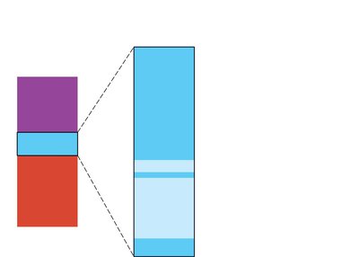
Blood is a fluid connective tissue essential for transporting substances throughout the body. It consists of two main components: plasma and formed elements (erythrocytes, leukocytes, and platelets).
Plasma: The liquid portion, making up about 55% of blood volume, contains water, proteins, nutrients, electrolytes, gases, and hormones.
Formed Elements: Includes erythrocytes (RBCs), leukocytes (WBCs), and platelets.
Hematocrit is the percentage of blood volume occupied by RBCs, typically 47% ± 5% in males and 42% ± 5% in females.
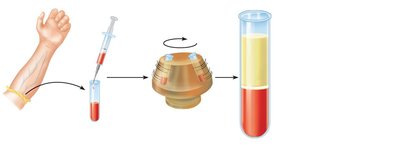
Blood is a sticky, opaque fluid with a pH of 7.35–7.45 and a temperature of about 38°C. It constitutes approximately 8% of body weight, with average volumes of 5–6 L for males and 4–5 L for females.
Functions of Blood
Blood performs three primary functions:
Distribution: Transports oxygen, nutrients, metabolic wastes, and hormones.
Regulation: Maintains body temperature, pH balance, and fluid volume.
Protection: Prevents blood loss (clotting) and defends against infection (immune cells and proteins).
Blood Plasma
Composition of Plasma
Plasma is 90% water and contains proteins (mainly produced by the liver), metabolic by-products, nutrients, electrolytes, respiratory gases, and hormones.
Proteins: 60% albumin, 36% globulins, 4% fibrinogen.
Nutrients: Glucose, amino acids, carbohydrates.
Electrolytes: Na+, K+, Ca2+, Cl–, HCO3–.
Respiratory Gases: O2 and CO2.
Formed Elements of Blood
Overview of Formed Elements
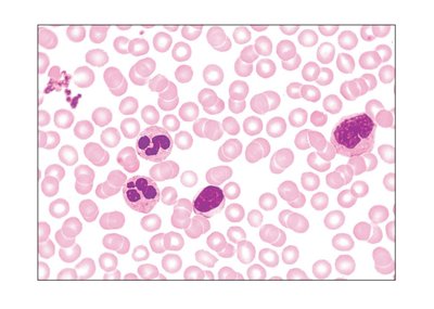
Formed elements include erythrocytes, leukocytes, and platelets. Only WBCs are complete cells; RBCs lack nuclei and organelles, and platelets are cell fragments. Most formed elements originate in bone marrow and have limited lifespans.
Erythrocytes (Red Blood Cells)
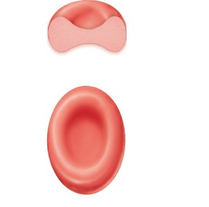
Erythrocytes are biconcave, anucleate cells specialized for gas transport. Their shape increases surface area for efficient gas exchange and provides flexibility.
Structure: Biconcave disc, no nucleus or organelles, filled with hemoglobin.
Function: Transport oxygen and carbon dioxide.
ATP Production: Anaerobic, so no oxygen is consumed by RBCs.
Hemoglobin Structure and Function
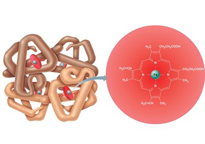
Hemoglobin is the protein responsible for oxygen transport. It consists of four globin chains (two alpha, two beta) and four heme groups, each containing an iron atom that binds oxygen.
O2 Loading: In lungs, forms oxyhemoglobin (ruby red).
O2 Unloading: In tissues, forms deoxyhemoglobin (dark red).
CO2 Transport: Forms carbaminohemoglobin, carrying 20% of CO2 in blood.
Hematopoiesis and Erythropoiesis
Hematopoiesis
Hematopoiesis is the process of blood cell formation, occurring in red bone marrow. Hemocytoblasts (hematopoietic stem cells) give rise to all formed elements.
Erythropoiesis
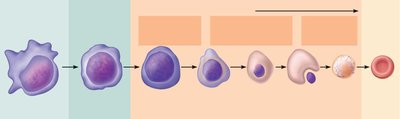
Erythropoiesis is the production of RBCs. It involves several developmental stages:
Transformation of hemocytoblast to proerythroblast
Ribosome synthesis
Hemoglobin accumulation
Ejection of nucleus, formation of reticulocyte, then mature erythrocyte
Regulation of Erythropoiesis
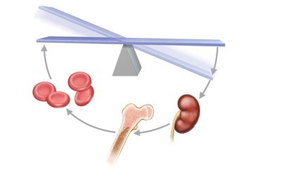
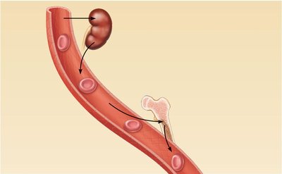
RBC production is regulated by hormonal controls (mainly erythropoietin, EPO) and adequate supplies of iron, amino acids, and B vitamins. EPO is released by the kidneys in response to hypoxia.
Fate and Destruction of Erythrocytes
Lifecycle of RBCs
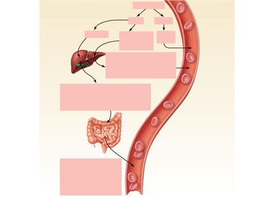
RBCs have a lifespan of 100–120 days. Old RBCs are removed by macrophages in the spleen, and their components are recycled.
Iron: Salvaged and reused.
Heme: Degraded to bilirubin, excreted in bile and feces.
Globin: Metabolized into amino acids.
Erythrocyte Disorders
Anemia
Anemia is a condition of abnormally low oxygen-carrying capacity in the blood, resulting in fatigue, paleness, shortness of breath, and chills. Causes include:
Insufficient erythrocytes: Hemorrhagic, hemolytic, or aplastic anemia.
Low hemoglobin content: Iron-deficiency or pernicious anemia.
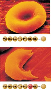
Abnormal hemoglobin: Thalassemias and sickle-cell anemia.
Polycythemia
Polycythemia is an excess of RBCs, increasing blood viscosity. It may result from bone marrow cancer, high altitude, or blood doping.
Leukocytes (White Blood Cells)
Types and Functions
Leukocytes make up less than 1% of blood volume and are crucial for immune defense. They can leave capillaries and move through tissues by diapedesis and chemotaxis.
Granulocytes
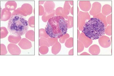
Granulocytes include neutrophils, eosinophils, and basophils. They have lobed nuclei and cytoplasmic granules.
Neutrophils: Most numerous, phagocytic, "bacteria slayers".
Eosinophils: Digest parasitic worms, modulate immune response.
Basophils: Rarest, contain histamine, functionally similar to mast cells.
Agranulocytes
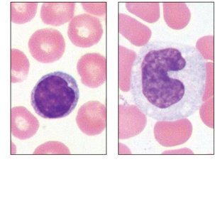
Agranulocytes lack visible granules and include lymphocytes and monocytes.
Lymphocytes: Crucial to immunity; T cells attack infected cells, B cells produce antibodies.
Monocytes: Largest WBCs, differentiate into macrophages, phagocytic.
Summary Table of Formed Elements
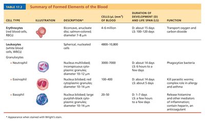
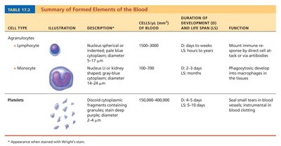
The following tables summarize the main formed elements of blood, their characteristics, and functions:
Cell Type | Description | Cells/μL | % of Blood | Duration of Development & Life Span | Function |
|---|---|---|---|---|---|
Erythrocytes | Biconcave, anucleate disc; salmon-colored | 4-6 million | ~45% | About 15 days; 100-120 days | Transport oxygen and carbon dioxide |
Neutrophil | Nucleus multilobed; pale red and blue granules | 3000-7000 | 50-70% | About 14 days; 6 hours | Phagocytize bacteria |
Eosinophil | Nucleus bilobed; red granules | 100-400 | 2-4% | About 14 days; 5 days | Kill parasitic worms; allergy and asthma |
Basophil | Nucleus bilobed; large blue-purple granules | 20-50 | 0.5-1% | About 1-7 days; few hours-days | Release histamine; contain heparin |
Cell Type | Description | Cells/μL | % of Blood | Duration of Development & Life Span | Function |
|---|---|---|---|---|---|
Lymphocyte | Nucleus spherical or indented; pale blue cytoplasm | 1500-3000 | 25-45% | Days to weeks; hours to years | Mount immune response |
Monocyte | Nucleus U or kidney-shaped; gray-blue cytoplasm | 100-700 | 3-8% | 2-3 days; months | Phagocytosis; develop into macrophages |
Platelets | Discoid cytoplasmic fragments; deep purple granules | 150,000-400,000 | <1% | 4-5 days; 5-10 days | Seal small tears in blood vessels; blood clotting |
Platelets and Hemostasis
Platelet Function
Platelets are cell fragments essential for blood clotting. They form temporary plugs at injury sites and release chemicals to promote clotting.
Hemostasis
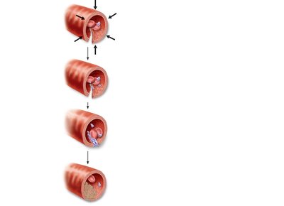
Hemostasis is the process of stopping bleeding, involving three steps:
Vascular spasm: Vasoconstriction of damaged vessel.
Platelet plug formation: Platelets adhere to exposed collagen, become sticky, and release messengers.
Coagulation: Fibrin forms a mesh, trapping blood cells and platelets to form a clot.
Coagulation
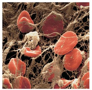
Coagulation transforms blood from a liquid to a gel, reinforcing the platelet plug with fibrin threads. The process involves:
Formation of prothrombin activator (intrinsic and extrinsic pathways)
Conversion of prothrombin to thrombin
Thrombin catalyzes joining of fibrinogen to form a fibrin mesh
Blood Groups and Transfusion
ABO Blood Groups
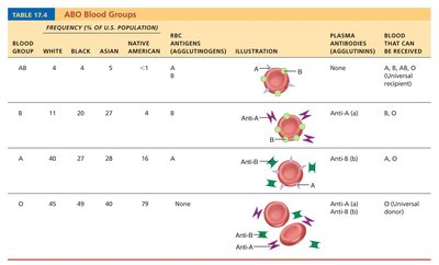
Blood types are classified based on the presence of agglutinogens (A and B) on RBC surfaces. The four main types are A, B, AB, and O. Plasma contains antibodies (agglutinins) against antigens not present on the individual's RBCs.
Blood Group | RBC Antigens | Plasma Antibodies | Blood That Can Be Received |
|---|---|---|---|
AB | A, B | None | A, B, AB, O (Universal recipient) |
B | B | Anti-A | B, O |
A | A | Anti-B | A, O |
O | None | Anti-A, Anti-B | O (Universal donor) |
Blood Typing
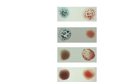
Blood typing involves mixing blood with anti-A and anti-B sera. Agglutination indicates the presence of the corresponding antigen.
Diagnostic Blood Tests
Common Tests
Diagnostic tests include hematocrit, blood glucose, microscopic examination of RBCs, differential WBC count, prothrombin time, platelet counts, and complete blood count (CBC).
Summary
Blood is a complex tissue with critical roles in transport, regulation, and protection. Understanding its composition, function, and disorders is essential for clinical practice and physiology.
