 뒤로
뒤로Blood Vessels and Circulation: Structure, Function, and Regulation
스터디 가이드 - 스마트 노트
 자료에 맞춘 맞춤형 노트, 핵심 정의, 예시, 맥락을 확장해 제공합니다.
Blood Vessels and Circulation
Overview of Blood Vessel Classes
The circulatory system is composed of five general classes of blood vessels, each with distinct structural and functional characteristics. Understanding these classes is essential for grasping how blood is transported throughout the body.
Arteries: Carry blood away from the heart under high pressure.
Arterioles: Small branches of arteries that regulate blood flow into capillaries.
Capillaries: Microscopic vessels where exchange of gases, nutrients, and waste occurs.
Venules: Small vessels that collect blood from capillaries and transport it to veins.
Veins: Return blood to the heart under lower pressure; often contain valves to prevent backflow.
Example: The aorta is the largest artery, while the venae cavae are the largest veins.
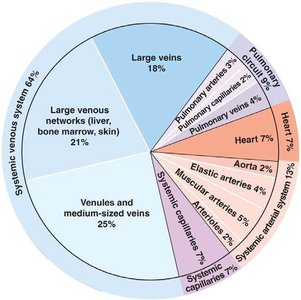
Distribution of Blood in the Body
Blood is unevenly distributed among the various vessel types, with the majority residing in the venous system. This distribution is crucial for understanding capacitance and the body's ability to mobilize blood during physiological demands.
Systemic venous system: Contains about 64% of total blood volume.
Large veins: 18%
Venules and medium-sized veins: 25%
Large venous networks: 21%
Arteries, capillaries, and heart: Smaller percentages, but critical for active circulation.
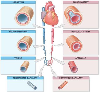
Types and Functions of Blood Vessels
Blood vessels are specialized for their roles in circulation. Their structure directly relates to their function, such as elasticity in arteries and permeability in capillaries.
Elastic arteries: Conduct blood and dampen pressure fluctuations.
Muscular arteries: Distribute blood to specific organs.
Arterioles: Control blood flow into capillary beds.
Capillaries: Exchange materials between blood and tissues.
Venules and veins: Collect blood and return it to the heart; veins have valves to prevent backflow.
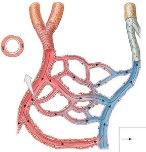
Capillary Function and Regulation
Capillaries are the primary site for exchange of gases, nutrients, and waste. Blood flow through capillary beds is regulated by precapillary sphincters, which respond to tissue oxygen demand.
High tissue demand for oxygen: Precapillary sphincters open, increasing blood flow.
Low tissue demand for oxygen: Precapillary sphincters close, reducing blood flow.
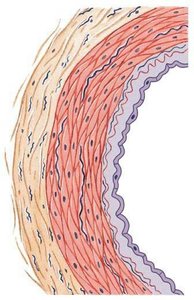
Structure of Blood Vessel Walls
Blood vessels have three distinct layers, each contributing to their function:
Tunica intima: Innermost layer, composed of endothelium.
Tunica media: Middle layer, primarily smooth muscle and elastic fibers; responsible for vasoconstriction and vasodilation.
Tunica externa: Outermost layer, connective tissue providing structural support.
Special Features of Veins: Valves
Veins, especially in the limbs, contain valves that prevent the backflow of blood. This is essential for returning blood to the heart against gravity.
Valve closed: Prevents backflow.
Valve opened: Allows blood to move toward the heart.
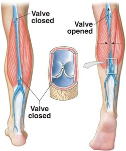
Physiology of Circulation: Blood Flow and Pressure
Blood flow is driven by pressure gradients and opposed by resistance. Understanding these relationships is key to cardiovascular physiology.
Blood flow (F): Directly proportional to pressure gradient (P), inversely proportional to resistance (R).
Key equations:
Example: Blood pressure is highest in arteries and lowest in veins.
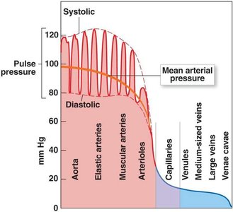
Factors Affecting Blood Pressure
Blood pressure is influenced by peripheral resistance, cardiac output, and blood volume. Each factor can be modulated physiologically or pathologically.
Peripheral resistance (PR): Opposition to blood flow; increases with vessel constriction, length, and blood viscosity.
Cardiac output (CO): Volume of blood pumped by the heart per minute; increases with heart rate and stroke volume.
Blood volume: More blood increases stroke volume and cardiac output, raising blood pressure.
Example: Vasoconstriction increases PR and BP; vasodilation decreases PR and BP.
Vessel Properties in Circulation
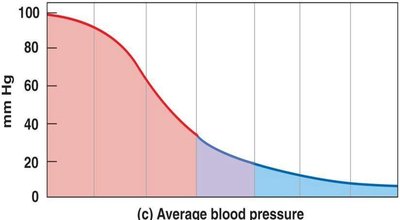
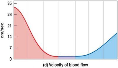
Vessel diameter, cross-sectional area, blood pressure, and blood velocity vary across the circulatory system, affecting how blood is transported and exchanged.
Vessel diameter: Largest in the aorta and venae cavae.
Cross-sectional area: Greatest in capillaries, facilitating exchange.
Blood pressure: Highest in arteries, lowest in veins.
Blood velocity: Fastest in arteries, slowest in capillaries.
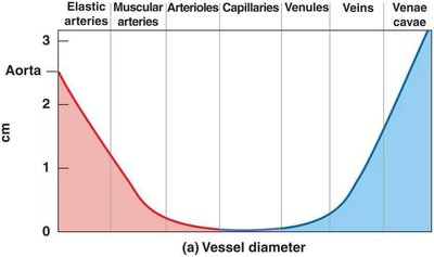
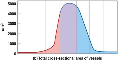
Regulation of Blood Pressure
Blood pressure is regulated by local, neural, and hormonal mechanisms. These ensure adequate perfusion of tissues under varying conditions.
Autoregulation: Local control within capillary beds; responds to changes in O2, CO2, K+, H+, histamines, and temperature.
Central Nervous System: Cardiac and vasomotor centers in the medulla oblongata adjust cardiac output and peripheral resistance.
Chemical and Hormonal Regulation: Vasoconstrictors (norepinephrine, angiotensin II, ADH) and vasodilators (NO, ANP) modulate vessel tone.
Baroreceptors: Detect changes in blood pressure and initiate reflexes to maintain homeostasis.
Chemoreceptors: Respond to changes in blood chemistry, influencing cardiovascular responses.
Example: During exercise, local vasodilators increase blood flow to active muscles.
Systemic vs. Pulmonary Circulation
The circulatory system is divided into systemic and pulmonary circuits, each serving distinct functions.
Systemic circuit: Delivers oxygenated blood to tissues and returns deoxygenated blood to the heart.
Pulmonary circuit: Carries deoxygenated blood to the lungs for gas exchange and returns oxygenated blood to the heart.
Example: The pulmonary arteries carry blood from the right ventricle to the lungs, while the aorta distributes blood from the left ventricle to the body.
Key Terms and Definitions
Artery: Vessel carrying blood away from the heart.
Arteriole: Small artery leading to capillaries.
Capillary: Microscopic vessel for exchange.
Venule: Small vein collecting blood from capillaries.
Vein: Vessel returning blood to the heart.
Fenestrated capillary: Capillary with pores for rapid exchange.
Continuous capillary: Capillary with uninterrupted endothelium.
Precapillary sphincter: Muscle regulating blood flow into capillaries.
Tunica intima, media, externa: Layers of vessel walls.
Valve: Structure preventing backflow in veins.
Blood pressure: Force exerted by blood on vessel walls.
Pulse: Rhythmic expansion of arteries.
Peripheral resistance: Resistance to blood flow in vessels.
Cardiac center: CNS region controlling heart rate.
Vasomotor center: CNS region controlling vessel tone.
Vasoconstrictors/vasodilators: Chemicals affecting vessel diameter.
Baroreceptor: Pressure sensor in vessels.
Chemoreceptor: Chemical sensor in vessels.
Pulmonary circuit: Blood flow to and from lungs.
Systemic circuit: Blood flow to and from body tissues.
Deep veins/superficial veins: Veins located deep or near the surface.
Additional info: These notes expand upon brief points in the original material, providing academic context and definitions for clarity.
