 뒤로
Bone Tissue and the Skeletal System: Structure, Function, and Development
스터디 가이드 - 스마트 노트
 자료에 맞춘 맞춤형 노트, 핵심 정의, 예시, 맥락을 확장해 제공합니다.
Bone Tissue Basics
Overview and Functions
Bone tissue, also known as osseous tissue, forms the hard, dense connective tissue of the adult skeleton. The skeletal system is composed of bones and cartilage, which are dynamic organs capable of growth, repair, and renewal. The adult human skeleton consists of 206 bones, each serving multiple essential functions:
Support: Provides a scaffold to support the body and its weight.
Movement: Serves as points of attachment for muscles, facilitating movement.
Protection: Shields internal organs (e.g., skull protects the brain, rib cage protects heart and lungs).
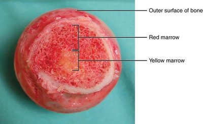
Blood Cell Production: Red marrow is involved in hematopoiesis, producing red and white blood cells and platelets.
Mineral and Fat Storage: Acts as a reservoir for calcium and phosphorus; yellow marrow stores triglycerides.
Organization of Bone Tissue
Types of Bone Cells
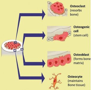
Bone tissue contains four main types of cells, each with distinct roles in bone formation, maintenance, and remodeling:
Osteogenic cells: Stem cells with a high mitotic index, found in deep layers of the periosteum and marrow. They differentiate into osteoblasts.
Osteoblasts: Responsible for forming bone matrix by synthesizing and secreting collagen and calcium salts. Located in growing portions of bone.
Osteocytes: Mature bone cells that maintain the mineral concentration of the matrix. Entrapped in lacunae within the calcified matrix.
Osteoclasts: Multinucleated cells that resorb bone, breaking down old or unneeded bone and releasing calcium into the blood.
General Structure of Bone Tissue
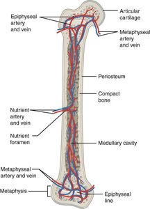
Bones are composed of a small number of cells embedded in a matrix of collagen fibers and inorganic crystals (hydroxyapatite). This structure provides both strength and flexibility. The periosteum covers the outer surface, while the endosteum lines the inner surfaces. Blood vessels and nerves penetrate bone to nourish and regulate growth.
Microscopic Organization of Bone Tissue
Lamellae, Trabeculae, and Waterways
Bone tissue is organized into lamellae (layers of calcified matrix) and trabeculae (lattice-like network in spongy bone). Spaces called lacunae contain osteocytes and extracellular fluid, while canaliculi are tiny canals that allow communication and nutrient transport between osteocytes.
Types of Bone Tissue
Compact vs. Spongy Bone
Most bones contain both compact and spongy bone tissue, with their distribution varying by function:
Compact bone: Dense and strong, arranged in osteons with concentric lamellae. Provides support and protection, found under the periosteum and in the diaphysis of long bones.
Spongy bone: Lattice-like network (trabeculae) that lightens bone and supports weight distribution. Houses red marrow for hematopoiesis.
Classification of Bones by Shape
Bone Shapes and Functions
Bones are classified by their shapes, which relate to their functions:
Long bones: Cylinder-like, longer than wide (e.g., femur, humerus). Provide leverage.
Short bones: Cube-like, equal in length, width, and thickness (e.g., carpals, tarsals). Provide stability and support.
Flat bones: Thin and curved (e.g., sternum, ribs, cranial bones). Protect internal organs.
Irregular bones: Complex shapes (e.g., vertebrae, facial bones). Protect internal organs.
Sesamoid bones: Small, round, embedded in tendons (e.g., patella). Protect tendons from compressive forces.
Gross Anatomy of a Long Bone
Parts and Coverings
Long bones have distinct anatomical regions:
Epiphyses: Ends of long bones, filled with spongy bone and red marrow.
Metaphysis: Region between diaphysis and epiphyses, contains epiphyseal plate/line.
Diaphysis: Tubular shaft, contains medullary cavity with yellow marrow.
Endosteum: Membranous lining for growth, repair, and remodeling.
Periosteum: Fibrous membrane covering outer surface, contains blood vessels and nerves.
Articular cartilage: Reduces friction at joints, acts as a shock absorber.
Bone Formation and Development
Ossification Processes
Bone formation (osteogenesis) begins in embryonic life and continues through adulthood. Two main processes are involved:
Intramembranous ossification: Bone develops directly from sheets of mesenchymal tissue. Forms facial bones, cranial bones, and clavicle.
Endochondral ossification: Bone develops from hyaline cartilage templates. Forms long bones and bones at the base of the skull.
Bone Growth
Appositional vs. Interstitial Growth
Bone grows in both diameter and length:
Appositional growth: Increase in diameter via osteoblasts beneath the periosteum and osteoclasts resorbing old bone lining the medullary cavity.
Interstitial growth: Increase in length via cartilage formation at the epiphyseal plate and ossification at the diaphyseal side.
Epiphyseal Plate and Zones
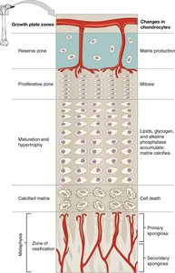
The epiphyseal plate is a longitudinal growth zone between the epiphysis and metaphysis. It consists of several zones:
Reserve zone: Small chondrocytes anchor the plate to the epiphysis.
Proliferative zone: Stacks of chondrocytes produced for growth.
Zone of maturation and hypertrophy: Older, larger chondrocytes contribute to growth.
Zone of calcified matrix: Dead chondrocytes due to calcification; capillaries and osteoblasts form bone tissue.
Bone Health and Remodeling
Role of Exercise
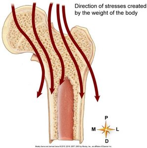
Regular exercise, especially resistance training, stimulates bone tissue deposition, increasing bone strength and density. Mechanical stress is essential for bone health, while lack of stress leads to bone loss and increased risk of osteoporosis.

Osteoporosis
Osteoporosis is a condition characterized by decreased bone mass and increased fragility. It results from an imbalance between bone resorption and deposition, often due to aging, hormonal changes, or lack of mechanical stress.
Calcium Homeostasis and Hormonal Regulation
Calcium Regulation
Calcium is the most abundant mineral in the body, with 99% stored in bones. It is essential for bone mineralization, muscle contraction, blood coagulation, and nerve conduction. Calcium homeostasis is regulated by hormones:
Calcitonin: Released from the thyroid gland when blood calcium is high; stimulates osteoblasts and inhibits osteoclasts.
Parathyroid hormone (PTH): Released from parathyroid glands when blood calcium is low; stimulates osteoclasts, increases renal absorption, and promotes vitamin D synthesis.
Calcitriol: Active form of vitamin D; increases calcium and phosphate absorption from the digestive tract.
Growth hormone: Increases length and mineralization of bones.
Thyroid hormones: Promote osteoblast activity and bone matrix synthesis.
Sex hormones: Promote osteoblast activity during adolescent growth and help convert epiphyseal plate to epiphyseal line.
Summary Table: Bone Cell Functions
Cell Type | Function |
|---|---|
Osteogenic cell | Stem cell; differentiates into osteoblasts |
Osteoblast | Forms bone matrix |
Osteocyte | Maintains bone tissue |
Osteoclast | Resorbs bone |
Additional info: These notes expand on brief points with academic context, definitions, and examples to provide a comprehensive mini-study guide for exam preparation in Anatomy & Physiology.
