뒤로
Bones and Bone Tissue: Structure, Function, and Growth
스터디 가이드 - 스마트 노트
 자료에 맞춘 맞춤형 노트, 핵심 정의, 예시, 맥락을 확장해 제공합니다.
Bones and Bone Tissue
Overview of the Skeletal System
The skeletal system consists of bones, joints, and their associated supporting tissues. Bones are the main organs of this system, composed of dense collagenous connective tissue, osseous tissue, and bone marrow. The skeletal system provides structural support, protection, and facilitates movement.
Functions of the Skeletal System
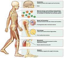
Bones serve several essential functions in the human body:
Protection: Guards vital organs such as the brain and heart.
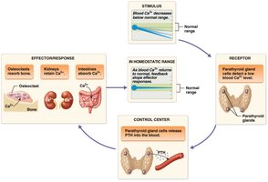
Mineral Storage and Acid-Base Homeostasis: Stores minerals (e.g., Ca2+, PO43-) necessary for electrolyte and acid-base balance.
Blood Cell Formation: Red bone marrow is the site of hematopoiesis (blood cell production).
Fat Storage: Yellow bone marrow stores triglycerides.
Movement: Muscles attach to bones, enabling movement.
Support: Provides structural support for the body.
Bone Structure
Classification of Bones by Shape
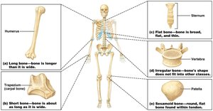
Bones are classified into five categories based on their shape:
Long Bones: Longer than they are wide (e.g., humerus).
Short Bones: About as long as they are wide (e.g., trapezium).
Flat Bones: Broad, flat, and thin (e.g., sternum).
Irregular Bones: Shape does not fit other classes (e.g., vertebra).
Sesamoid Bones: Round, flat bones found within tendons (e.g., patella).
Structure of a Long Bone
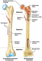
Long bones have a distinct anatomy that supports their function:
Diaphysis: Shaft of the bone.
Epiphysis: Ends of the bone.
Epiphyseal Line: Remnant of the epiphyseal plate (growth area).
Medullary Cavity: Central cavity containing bone marrow.
Periosteum: Outer membrane covering the bone.
Endosteum: Lining of the internal bone surfaces.
Compact Bone: Dense outer layer.
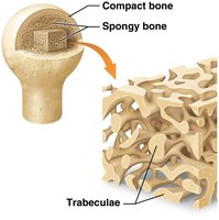
Spongy Bone: Inner layer with trabeculae.
Structure of Other Bones
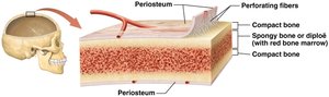
Other bone types (short, flat, irregular, sesamoid) lack diaphyses, epiphyses, medullary cavities, epiphyseal lines, or plates. They possess periosteum, perforating fibers, blood vessels, and nerves. The outer layer is compact bone, with spongy bone in the middle. Skull bones may contain sinuses.
Bone Marrow
Bone marrow exists in two forms:
Red Bone Marrow: Contains hematopoietic cells for blood formation. More abundant in children; in adults, found in pelvis, proximal femur and humerus, vertebrae, ribs, sternum, clavicles, scapulae, and some skull bones.
Yellow Bone Marrow: Contains triglycerides, blood vessels, and adipocytes. Increases with age.
Microscopic Structure of Bone
Extracellular Matrix (ECM) of Bone
The bone ECM is divided into inorganic and organic components:
Inorganic Matrix: Makes up 65% of bone's weight. Contains minerals such as calcium and phosphorus, stored as hydroxyapatite crystals (). Also stores bicarbonate, potassium, magnesium, and sodium.
Organic Matrix: Makes up 35% of bone's weight. Primarily collagen, which forms cross-links to resist torsion and tensile forces.
Bone Cells
Three main types of bone cells are involved in bone formation and maintenance:
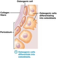
Osteogenic Cells: Stem cells that differentiate into osteoblasts when stimulated.
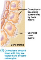
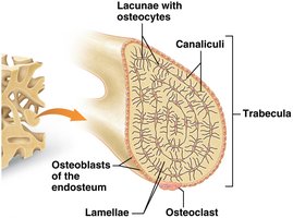
Osteoblasts: Metabolically active cells in periosteum and endosteum; secrete organic matrix and are responsible for bone deposition.
Osteocytes: Mature bone cells that have surrounded themselves with bone matrix in lacunae; maintain bone tissue but do not actively synthesize matrix.
Osteoclasts: Large multinucleated cells responsible for bone resorption; secrete hydrogen ions and enzymes to break down bone matrix and release minerals into the blood.
Histology of Bone
Bone tissue is organized into two main types: compact and spongy bone.
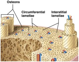
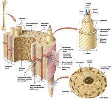
Compact Bone: Composed of osteons (Haversian systems), which are concentric rings (lamellae) surrounding a central canal containing blood vessels and nerves. Osteocytes reside in lacunae, connected by canaliculi for communication and resource exchange. Osteons are not permanent; they are remodeled by osteoclasts and osteoblasts.
Interstitial Lamellae: Fill spaces between osteons; remnants of old osteons.
Circumferential Lamellae: Outer and inner layers just inside periosteum.
Perforating Canals (Volkmann’s Canals): Originate from blood vessels in periosteum and run perpendicular to central canals.
Spongy Bone: Less densely packed, not weight-bearing, provides protective structure for bone marrow. Composed of trabeculae, which are bony branches covered with endosteum and contain concentric lamellae with lacunae between.
Ossification (Bone Formation)
Types of Ossification
Ossification, or osteogenesis, is the process of bone formation during the embryonic period and childhood. Most bones are fully formed by age 7. There are two main processes:
Intramembranous Ossification: Bone develops from a membrane of embryonic connective tissue. Occurs in skull and clavicles. Larger bones have multiple primary ossification centers; pieces fuse over time. Fontanels (soft spots) are present in infants.
Endochondral Ossification: Bone develops from a model of hyaline cartilage. Occurs in all bones below the head except clavicles.
Bone Growth
Longitudinal Growth
Bone length increases through division of chondrocytes in the epiphyseal plate, located at the epiphysis closest to the diaphysis. Growth occurs in distinct zones:
Zone of Reserve Cartilage: Anchoring layer closest to epiphysis.
Zone of Proliferation: Actively dividing chondrocytes.
Zone of Hypertrophy and Maturation: Mature chondrocytes.
Zone of Calcification: Dead chondrocytes, some calcified.
Zone of Ossification: Calcified chondrocytes and osteoblasts.
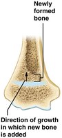
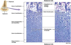
Longitudinal growth continues as long as mitosis occurs in the zone of proliferation. Growth slows around ages 12–15, and the epiphyseal plate eventually ossifies, forming the epiphyseal line and stopping further lengthening.
Appositional Growth
Appositional growth increases bone width. Osteoblasts between the periosteum and bone surface lay down new bone, forming new circumferential lamellae. Older lamellae are removed or restructured into osteons. Width increase continues even after epiphyseal plates have closed.
Disorders Associated with Bone Growth
Achondroplasia: Most common cause of dwarfism; gene defect affects growth factor receptor on cartilage, leading to short limbs and facial abnormalities.
Gigantism: Excess growth hormone in childhood causes abnormal height.
Acromegaly: Excess growth hormone in adulthood causes enlargement of bone, cartilage, and soft tissue.
Pituitary Dwarfism: Too little growth hormone results in short stature.
Hormonal Regulation of Bone Growth
Growth Hormone (GH): Increases cell division in nearly all tissues; affects both longitudinal and appositional growth.
Testosterone: Increases appositional growth, rate of mitosis in epiphyseal plate, and accelerates closure of epiphyseal plate.
Estrogen: Increases rate of longitudinal bone growth, inhibits osteoclast activity, and accelerates closure of epiphyseal plate faster than testosterone.
Bone Remodeling
Process and Purpose
Bone remodeling is the continuous process of bone deposition and resorption, maintaining calcium ion homeostasis and replacing old bone with new. It adapts bone to tension and stress, and repairs brittle bone.
In adults: Bone formation and loss occur simultaneously.
In children: Deposition occurs at a much faster rate than resorption.
Factors Affecting Remodeling
Mechanical Stress: Compression stimulates deposition; tension stimulates deposition; continuous pressure stimulates resorption.
Hormones: Various hormones regulate remodeling.
Age: Remodeling rate changes with age.
Calcium and Vitamin Intake: Calcium, vitamin D, C, K, and protein are essential for proper remodeling.
Bone Repair
Fracture Types
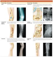
Fractures are breaks in bone and are classified as:
Simple Fractures: Skin and tissue around fracture remain intact.
Compound Fractures: Skin and tissues around fracture are damaged.
Bone Repair Process
Bone repair involves several steps:
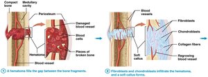
A hematoma fills the gap between bone fragments.
Fibroblasts and chondroblasts form a soft callus.
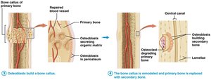
Osteoblasts build a bone callus of primary bone.
The bone callus is remodeled and replaced with secondary bone.