 뒤로
뒤로Bones and Skeletal Tissues: Structure, Function, and Microscopic Anatomy
스터디 가이드 - 스마트 노트
 자료에 맞춘 맞춤형 노트, 핵심 정의, 예시, 맥락을 확장해 제공합니다.
Bones and Skeletal Tissues
Introduction
The skeletal system provides the framework for the human body, supporting movement, protecting organs, and serving as a reservoir for minerals and blood cell production. Understanding the structure and function of bones and skeletal tissues is essential for comprehending human anatomy and physiology.
Skeletal Cartilages
Basic Structure, Types, and Locations
Skeletal cartilage is a resilient, molded tissue primarily composed of water, lacking blood vessels and nerves.
Perichondrium is a dense connective tissue layer surrounding cartilage, providing nutrients and resisting outward expansion.
Cartilage cells, called chondrocytes, reside in lacunae within a jelly-like extracellular matrix.
Types of Cartilage
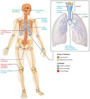
Hyaline cartilage: Most abundant; provides support, flexibility, and resilience. Found in articular surfaces, costal cartilage, respiratory structures, and nasal cartilage.
Elastic cartilage: Contains elastic fibers; found in the external ear and epiglottis.
Fibrocartilage: Contains thick collagen fibers; provides tensile strength. Located in menisci of the knee and intervertebral discs.
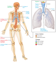
Growth of Cartilage
Appositional growth: New matrix is secreted against the external face of existing cartilage by cells in the perichondrium.
Interstitial growth: Chondrocytes divide and secrete new matrix from within the cartilage.
Calcification: Occurs during normal bone growth and aging; calcified cartilage is not bone.
Functions of Bones
Major Functions
Support: Framework for the body and soft organs.
Protection: Shields the brain, spinal cord, and vital organs.
Anchorage: Acts as levers for muscle action.
Mineral and growth factor storage: Reservoir for calcium, phosphorus, and growth factors.
Blood cell formation: Hematopoiesis in red marrow cavities.
Triglyceride storage: Fat storage in bone cavities for energy.
Hormone production: Osteocalcin regulates insulin secretion, glucose levels, and metabolism.
Classification of Bones
By Location
Axial skeleton: Skull, vertebral column, rib cage (long axis of body).
Appendicular skeleton: Limbs and girdles attaching limbs to axial skeleton.
By Shape
Long bones: Longer than wide (e.g., limb bones).
Short bones: Cube-shaped (wrist, ankle); sesamoid bones form within tendons (e.g., patella).
Flat bones: Thin, flat, slightly curved (sternum, scapulae, ribs, skull bones).
Irregular bones: Complicated shapes (vertebrae, hip bones).
Bone Structure
Gross Anatomy
Bones are organs containing bone tissue, nervous tissue, cartilage, fibrous connective tissue, muscle cells, and epithelial cells in blood vessels.
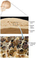
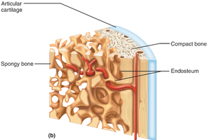
Two main types of bone tissue: compact bone (dense outer layer) and spongy bone (honeycomb of trabeculae).
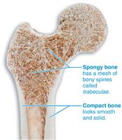
Structure of Short, Irregular, and Flat Bones
Thin plates of spongy bone (diploë) covered by compact bone.
Periosteum covers outside; endosteum covers inside.
Bone marrow is scattered throughout spongy bone; no defined marrow cavity.
Hyaline cartilage covers areas involved in movable joints.
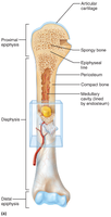
Structure of a Typical Long Bone
Diaphysis: Shaft; compact bone surrounds medullary cavity (yellow marrow in adults).
Epiphyses: Bone ends; compact bone externally, spongy bone internally; articular cartilage covers joint surfaces.
Epiphyseal line: Remnant of growth plate between diaphysis and epiphysis.
Membranes
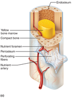
Periosteum: Double-layered membrane covering external bone surfaces (except joints); outer fibrous layer (dense irregular connective tissue), inner osteogenic layer (osteoprogenitor cells).
Contains nerve fibers, blood vessels, and perforating fibers anchoring periosteum to bone.
Endosteum: Delicate membrane covering internal bone surfaces, trabeculae, and canals; contains osteogenic cells.
Blood Vessels and Nerves
Bones are well vascularized; nutrient arteries and veins enter through nutrient foramina.
Epiphyseal arteries and veins supply the epiphyses; nerves accompany blood vessels.
Hematopoietic Tissue in Bones
Red marrow: Found in trabecular cavities of spongy bone and diploë of flat bones (sternum, hip bone).
In newborns, all spongy bone and medullary cavities contain red marrow; in adults, mainly in flat bones and heads of femur/humerus.
Yellow marrow can revert to red marrow if necessary (e.g., anemia).
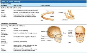
Bone Markings
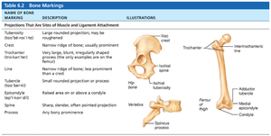
Serve as sites for muscle, ligament, and tendon attachment, joint formation, or passageways for blood vessels and nerves.
Three types: projections (attachment sites), surfaces (form joints), depressions/openings (passageways).
Name of Bone Marking | Description | Illustrations |
|---|---|---|
Crest | Narrow ridge of bone; usually prominent | Example: Iliac crest |
Trochanter | Very large, blunt, irregularly shaped process | Example: Femur |
Line | Narrow ridge of bone; less prominent than a crest | Example: Femur |
Tubercle | Small rounded projection or process | Example: Humerus |
Process | Any bony prominence | Example: Vertebra |
Name of Bone Marking | Description | Illustrations |
|---|---|---|
Head | Bony expansion carried on a narrow neck | Example: Femur head |
Facet | Smooth, nearly flat articular surface | Example: Vertebra |
Condyle | Rounded articular projection | Example: Mandible |
Foramen | Round or oval opening through a bone | Example: Skull |
Fossa | Shallow basin-like depression in a bone | Example: Skull |
Microscopic Anatomy of Bone
Bone Cells
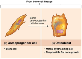
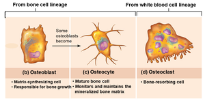
Osteoprogenitor cells: Stem cells in periosteum and endosteum; differentiate into osteoblasts or bone-lining cells.
Osteoblasts: Bone-forming cells; secrete osteoid (unmineralized bone matrix composed of collagen and calcium-binding proteins).
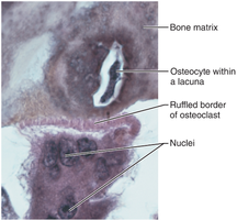
Osteocytes: Mature bone cells in lacunae; maintain bone matrix and act as stress sensors.
Bone-lining cells: Flat cells on bone surfaces; help maintain matrix.
Osteoclasts: Multinucleate cells derived from hematopoietic stem cells; responsible for bone resorption.
Microscopic Anatomy of Compact Bone
Osteon (Haversian system): Structural unit; elongated cylinder with concentric lamellae (rings of bone matrix).
Lamellae have collagen fibers running in alternating directions, providing strength and resistance to twisting.
Central (Haversian) canal: Runs through osteon core; contains blood vessels and nerves.
Perforating (Volkmann's) canals: Connect blood vessels and nerves of periosteum, medullary cavity, and central canal.
Lacunae: Small cavities containing osteocytes.
Canaliculi: Hairlike canals connecting lacunae; allow communication and nutrient/waste exchange between osteocytes.
Interstitial lamellae: Fill gaps between osteons or are remnants of old osteons.
Circumferential lamellae: Extend around the entire diaphysis; resist twisting of the long bone.
Microscopic Anatomy of Spongy Bone
Composed of trabeculae aligned along lines of stress, providing strength without weight.
Trabeculae contain irregularly arranged lamellae and osteocytes interconnected by canaliculi.
Capillaries in endosteum supply nutrients.
Chemical Composition of Bone
Organic Components
Include bone cells (osteoprogenitor cells, osteoblasts, osteocytes, bone-lining cells, osteoclasts) and osteoid (one-third of matrix).
Osteoid consists of ground substance and collagen fibers, providing tensile strength and flexibility.
Resilience is due to sacrificial bonds in collagen molecules that dissipate energy and prevent fractures.
Inorganic Components
Hydroxyapatites (mineral salts): Make up 65% of bone by mass; mainly tiny calcium phosphate crystals in and around collagen fibers.
Responsible for bone hardness and resistance to compression.
Mineral composition allows bones to last long after death, providing archaeological information.
