 뒤로
Cardiovascular System: Structure and Function Study Guide
스터디 가이드 - 스마트 노트
 자료에 맞춘 맞춤형 노트, 핵심 정의, 예시, 맥락을 확장해 제공합니다.
Cardiovascular System
I. Heart
The heart is a muscular organ located in the mediastinum, responsible for pumping blood throughout the body. It is suspended at its base by the great vessels and has distinct surfaces and borders.
Location: Mediastinum, anchored by great vessels.
Surfaces and Borders: Includes apex, base, diaphragmatic surface, sternocostal surface, left and right borders.
Surface Markings: Atrioventricular groove (coronary sulcus), anterior and posterior interventricular sulci.
Pericardium
The pericardium is a double-layered sac surrounding the heart and great vessels, providing protection and anchorage.
Fibrous Pericardium: Anchors heart, prevents overfilling.
Serous Pericardium: Consists of parietal and visceral layers, with the pericardial cavity between them filled with serous fluid.
Chambers of the Heart
Right Atrium: Contains pectinate muscles, right auricle, openings for superior/inferior vena cava and coronary sinus.
Interatrial Septum: Features fossa ovalis, a remnant of fetal foramen ovale.
Left Atrium: Contains left auricle.
Right Ventricle: Trabeculae carneae, papillary muscles (septal, anterior, posterior), moderator band, chordae tendineae, tricuspid valve, pulmonary semilunar valve.
Interventricular Septum: Separates ventricles.
Left Ventricle: Trabeculae carneae, papillary muscles (anterolateral, posteromedial), chordae tendineae, bicuspid/mitral valve, aortic semilunar valve.
Great Vessels
Systemic Pump: Ascending aorta, aortic arch (brachiocephalic, left common carotid, left subclavian arteries), superior and inferior vena cava.
Pulmonary Pump: Pulmonary trunk, pulmonary arteries, pulmonary veins.
Ligamentum Arteriosum: Remnant of fetal ductus arteriosus.
Coronary Circulation
Coronary arteries supply the heart muscle. Coronary dominance refers to which artery supplies the posterior-inferior wall.
Right-dominant: RCA gives rise to PDA (70-80%).
Left-dominant: LCx gives rise to PDA (5-10%).
Codominant: Both RCA and LCx contribute (10-20%).
Coronary Arteries: Left (LAD, LCx, left marginal, posterior left ventricular, PDA), Right (right marginal, PDA, posterior left ventricular).
Cardiac Veins: Great, middle, small cardiac veins, posterior vein of left ventricle, all drain into coronary sinus.
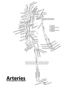
II. Arteries
Arteries carry blood away from the heart under high pressure. They have thick, tube-like walls and are paler than veins. Arteriosclerosis is the hardening and thickening of arterial walls.
Head and Neck: Aortic arch branches (brachiocephalic trunk, right/left subclavian, right/left common carotid), external/internal carotid, vertebral, internal thoracic, costocervical, thyrocervical, dorsal scapular arteries.
Upper Limb: Axillary, thoracoacromial, lateral thoracic, subscapular, humeral circumflex, brachial, deep brachial, radial, ulnar, palmar and dorsal arches, digital arteries.
Thoracic: Thoracic aorta, posterior intercostal arteries, internal thoracic artery.
Pelvic: Common iliac, internal/external iliac, gluteal, pudendal, obturator arteries.
Lower Limb: Femoral triangle (NAVeL mnemonic), femoral, deep femoral, circumflex femoral, popliteal, anterior/posterior tibial, fibular, plantar, dorsalis pedis arteries.
Abdominal: Abdominal aorta, celiac trunk (gastric, hepatic, splenic), mesenteric, renal, gonadal arteries.
Example: The femoral artery supplies blood to the lower limb and is a key landmark in the femoral triangle.
III. Veins
Veins return blood to the heart under low pressure. They are thinner, flatter, and darker than arteries. Many veins are paired with arteries (venae comitantes), aiding venous return.
Head and Neck: Superior vena cava, right/left brachiocephalic, internal/external jugular, subclavian veins.
Upper Limb: Axillary, brachial, basilic, cephalic, median cubital, median antebrachial veins.
Thoracic: Superior vena cava, azygos, hemiazygos, accessory hemiazygos, posterior intercostal veins.
Pelvic: Common iliac, external/internal iliac veins.
Lower Limb: Femoral, great saphenous veins.
Abdominal: Inferior vena cava, hepatic, hepatic portal, mesenteric, splenic, renal, suprarenal, gonadal veins.
Example: The hepatic portal vein collects nutrient-rich blood from the digestive organs and delivers it to the liver for processing.
IV. Clinical and Anatomical Notes
Arteriosclerosis: Hardening of arteries, especially coronary arteries, increases risk of vessel rupture.
Coronary Dominance: Important for understanding myocardial infarction risk and surgical planning.
Femoral Triangle: Key anatomical region for vascular access and clinical examination.
V. Summary Table: Major Arteries and Their Regions
Artery | Region Supplied |
|---|---|
Brachiocephalic trunk | Right head, neck, upper limb |
Left common carotid | Left head, neck |
Left subclavian | Left upper limb |
Femoral | Lower limb |
Renal | Kidneys |
Hepatic | Liver |
Coronary | Heart |
Superior mesenteric | Small intestine, colon |
Inferior mesenteric | Colon, rectum |
Additional info: The diagram included above is an idealized representation of the major arteries in the systemic circulation. Cadaveric variation is common, so anatomical study should account for individual differences.
