 뒤로
Central Nervous System: Structure, Function, and Pathways
스터디 가이드 - 스마트 노트
 자료에 맞춘 맞춤형 노트, 핵심 정의, 예시, 맥락을 확장해 제공합니다.
Central Nervous System
Ventricles of the Brain
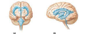
The brain contains four interconnected, fluid-filled chambers called ventricles. These spaces are filled with cerebrospinal fluid (CSF) and are essential for protecting and nourishing the brain.
Lateral Ventricles: Paired, C-shaped chambers located in each cerebral hemisphere.
Third Ventricle: Located in the midline, between the two halves of the diencephalon.
Fourth Ventricle: Located between the brainstem and the cerebellum.
Interventricular Foramen: Connects lateral ventricles to the third ventricle.
Cerebral Aqueduct: Connects the third ventricle to the fourth ventricle.
Central Canal: Extends from the fourth ventricle into the spinal cord.
Cerebrospinal Fluid (CSF)
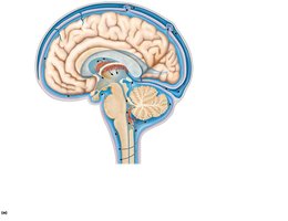
Cerebrospinal fluid is a clear, colorless liquid that surrounds the brain and spinal cord. It is produced by the choroid plexus and serves several important functions:
Bouyancy: Reduces the effective weight of the brain by 97%, preventing damage from its own mass.
Protection: Cushions the brain from physical shocks and jolts.
Chemical Stability: Nourishes neural tissue and removes waste, carrying chemical signals.
The choroid plexus is a cluster of capillaries enclosed by pia mater and surrounded by ependymal cells, responsible for CSF production.
CSF Circulation and Hydrocephalus
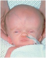
CSF is produced in the choroid plexus of each ventricle, circulates through the ventricles, enters the subarachnoid space via apertures, and is absorbed into the dural venous sinuses via arachnoid granulations. Disruption of CSF flow can lead to hydrocephalus, a condition characterized by abnormal accumulation of CSF.
Newborns: Unfused skull allows expansion, resulting in enlarged head.
Adults: Rigid skull increases intracranial pressure, risking brain damage.
Meninges of the Brain
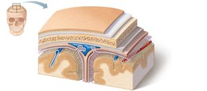
The meninges are three connective tissue membranes that cover the brain and spinal cord, providing protection and structural support:
Dura Mater: Tough, outermost layer with periosteal and meningeal layers; forms dural sinuses for venous drainage.
Arachnoid Mater: Middle layer; contains subarachnoid space filled with CSF and arachnoid granulations for CSF absorption.
Pia Mater: Delicate, innermost layer; adheres to brain surface and contains tiny blood vessels.
Functional Brain Systems
The Limbic System
The limbic system is a network of structures involved in emotion, motivation, and memory. It includes both diencephalic and cerebral components:
Amygdaloid Body (Amygdala): Processes fear and aggression.
Cingulate Gyrus: Coordinates emotional gestures and resolves mental conflicts.
Hippocampus: Essential for memory formation.
Other Structures: Dentate gyrus, parahippocampal gyrus, septal nuclei, and olfactory bulb.
Interaction with the frontal lobes allows emotional responses to conscious events. The limbic system's output travels through the hypothalamus, influencing autonomic and homeostatic functions.
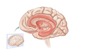
Reticular Formation
The reticular formation is a network of neurons in the brainstem that connects with the hypothalamus, thalamus, cerebral cortex, cerebellum, and spinal cord. It plays a crucial role in maintaining consciousness and filtering sensory input.
Reticular Activating System (RAS): Keeps the cerebral cortex alert and conscious.
Filtering: Filters out repetitive, familiar, or weak stimuli; only 1% of stimuli reach consciousness.
Inhibition: Can be inhibited, leading to coma.
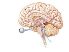
Higher Mental Functions
Language
Language processing occurs primarily in the association cortex of the left hemisphere, involving Broca’s area (speech production) and Wernicke’s area (language comprehension).
Aphasia: Language deficit due to lesions in Broca or Wernicke areas.
Nonfluent Aphasia: Lesion in Broca area; slow, labored speech.
Fluent Aphasia: Lesion in Wernicke area; speech is normal but nonsensical, with impaired comprehension.
Memory
Memory involves the storage and retrieval of information and is classified into several types:
Declarative: Facts and events.
Procedural: Skills and procedures.
Motor: Motor skills.
Emotional: Emotional experiences.
Memory processing involves stages:
Short-Term Memory (STM): Temporary storage, mainly in the hippocampus.
Long-Term Memory (LTM): Permanent storage, mainly in the cerebral cortex.
Factors Influencing Transfer: Excitement, rehearsal, association, and automatic memory.
Amnesia can be retrograde (loss of past memories) or anterograde (inability to form new memories).
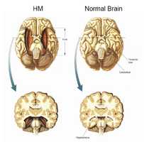
Brain Waves and EEG
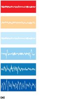
Brain waves reflect the electrical activity of the brain and are measured by an electroencephalogram (EEG). EEG is used to diagnose epilepsy, sleep disorders, brain lesions, and brain death.
Epilepsy: Characterized by abnormal electrical discharges.
Seizures: Include absence and tonic-clonic types.
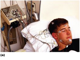
Consciousness
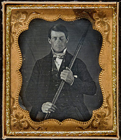
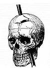
Consciousness encompasses perception, voluntary movement, cognition, memory, logic, and judgment. Famous cases, such as Phineas Gage, illustrate the effects of brain injury on personality and higher mental functions.
Sleep and Sleep-Wake Cycles
Sleep consists of two major types: Non-rapid eye movement (NREM) and Rapid eye movement (REM). NREM has four stages, while REM occurs about every 90 minutes and is associated with dreaming and increased vital signs.
Circadian Rhythms: Regulated by the suprachiasmatic nuclei of the hypothalamus.
Sleep Paralysis: Temporary inability to move during sleep transitions.
Narcolepsy: Caused by low orexin levels.
Insomnia: Associated with high orexin levels.
Importance: Sleep is restorative, helps analyze daily events, and eliminates unneeded synapses.

Spinal Cord Structure and Function
Gross Anatomy of the Spinal Cord
The spinal cord begins at the foramen magnum and ends at the L1 or L2 vertebra. Key features include:
Conus Medullaris: Tapered end of the spinal cord.
Cauda Equina: Bundle of spinal nerves below the conus medullaris.
Cervical and Lumbosacral Enlargements: Regions with increased neural input/output.
Two-way Communication: Between brain and body.
Reflex Center: Reflexes are initiated and completed at the spinal cord.
Spinal Cord Meninges
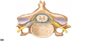
The spinal cord is protected by three meninges: dura mater, arachnoid mater, and pia mater. Spaces include the epidural (fat), subdural, and subarachnoid (CSF) spaces.
Spina Bifida
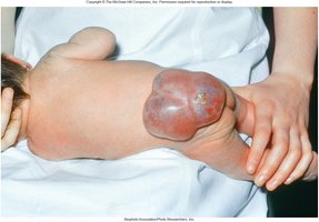
Spina bifida is a congenital defect where vertebrae fail to form a complete arch, exposing the spinal cord. It occurs in about 1 in 1,000 births and can be reduced by folic acid supplementation during pregnancy.
Spina bifida occulata: Mild form, minimal symptoms.
Spina bifida cystica: Severe form, visible cyst or sac.
Spinal Cord Gray Matter
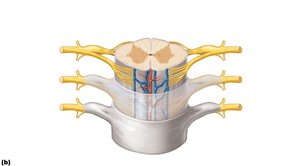
The spinal cord's gray matter is organized into horns:
Dorsal Horn: Contains interneurons receiving sensory input.
Ventral Horn: Contains motor neurons.
Lateral Horn: Contains autonomic motor neurons (in thoracic and upper lumbar regions).
Gray Commissure: Connects the two sides of gray matter.
Spinal Nerves
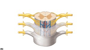
Spinal nerves are formed by the union of dorsal (sensory) and ventral (motor) roots. Dorsal root ganglia contain cell bodies of sensory neurons.
Spinal Cord White Matter
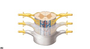
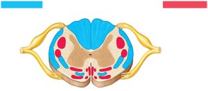
White matter in the spinal cord is organized into columns (funiculi): dorsal, ventral, and lateral. These columns contain ascending (sensory) and descending (motor) tracts.
Spinal Cord Pathways and Tracts
Spinal tracts are classified as ascending (sensory) or descending (motor). Four key features:
Decussation: Cross-over of fibers.
Relay: Chain of two or three neurons.
Somatotopy: Spatial relationship between CNS and body.
Symmetry: Paired pathways.
Ascending Pathways
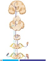
Ascending pathways transmit sensory information to the brain:
First Order Neuron: Sensory neuron from receptors to spinal cord/medulla.
Second Order Neuron: Interneuron from spinal cord/medulla to thalamus/cerebellum.
Third Order Neuron: Interneuron from thalamus to somatosensory cortex (not present in cerebellum).
Major pathways:
Medial Lemniscal Pathway: Discriminative touch, vibration, proprioception.
Spinothalamic Pathway: Pain, temperature, crude touch, pressure.
Spinocerebellar Tracts: Proprioception, muscle coordination.
Descending Pathways
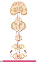
Descending pathways transmit motor commands from the brain to the body:
Direct Pathways: Originate in cerebral cortex; fast, fine movements (corticospinal tracts).
Indirect Pathways: Originate in brainstem; control axial muscles, balance, posture, coarse limb movements, and head/neck/eye movements.
Upper Motor Neuron: Begins in primary motor cortex or brainstem.
Lower Motor Neuron: Begins in ventral horn; innervates skeletal muscles.
Examples:
Corticospinal (Pyramidal) Tracts: Fast, fine movements.
Tectospinal Tract: Head movement in response to visual stimuli.
Rubrospinal Tract: Controls flexor muscles.
Vestibulospinal and Reticulospinal Tracts: Balance and posture.
 ----------------------------------------
