 뒤로
Central, Peripheral, and Autonomic Nervous Systems: Structure and Function
스터디 가이드 - 스마트 노트
 자료에 맞춘 맞춤형 노트, 핵심 정의, 예시, 맥락을 확장해 제공합니다.
Overview of the Brain
Main Regions of the Brain
The human brain is divided into several key regions, each with distinct functions essential for sensory processing, motor control, and integration of information.
Brainstem: Composed of the midbrain, pons, and medulla oblongata. It relays information between the brain and spinal cord and controls vital functions such as heartbeat and breathing.
Cerebellum: Coordinates movement, posture, and balance.
Cerebrum: Responsible for higher-order functions including perception, movement, and integration.
Diencephalon: Includes the thalamus, hypothalamus, and epithalamus, which regulate sensory relay, autonomic functions, and hormone secretion.
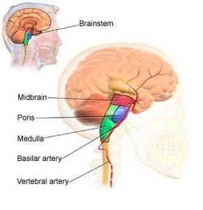
Brainstem Structure and Function
Medulla Oblongata: Relays sensory and motor signals; controls heart rate, breathing, and reflexes such as swallowing.
Pons: Connects cerebellum hemispheres and relays signals between medulla and midbrain; involved in breathing regulation.
Midbrain: Relays motor output and sensory input; controls eye movement and responses to auditory stimuli.
Protection: Meninges, Blood-Brain Barrier & Cerebral Spinal Fluid
Cranial Meninges
The brain and spinal cord are protected by three layers of connective tissue called meninges:
Dura Mater: Tough, outermost layer; in the cranium, it has two layers and forms extensions that separate brain regions.
Arachnoid Mater: Middle, web-like layer.
Pia Mater: Innermost layer; closely adheres to brain and spinal cord, providing vascular support.
Blood-Brain Barrier (BBB)
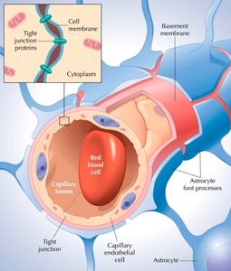
The BBB is a selective barrier formed by astrocytes and tight junctions between capillary endothelial cells. It protects the brain from harmful substances while allowing essential nutrients to pass.
Hydrophilic substances: Glucose (actively transported), creatinine, urea, ions (cross slowly).
Hydrophobic substances: Oxygen, carbon dioxide, alcohol, anesthetics (cross easily).
Proteins and antibiotics: Do not pass through the BBB.
Cerebral Spinal Fluid (CSF)
CSF is a clear, colorless fluid that protects the brain and spinal cord, maintains homeostasis, and circulates nutrients.
Production: Produced by choroid plexuses in ventricles, covered by ependymal cells.
Functions: Mechanical protection, chemical protection, circulation.
Composition: Glucose, proteins, lactic acid, urea, ions, white blood cells.
Circulation Pathway: Lateral ventricles → third ventricle → cerebral aqueduct → fourth ventricle → subarachnoid space → reabsorbed via arachnoid villi.
Cerebellum
Location & Composition
The cerebellum is located at the posterior, inferior aspect of the cranium and is the second largest brain region.
Transverse fissure: Separates cerebellum from cerebrum.
Vermis: Central constricted area.
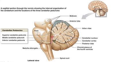
Cerebellar hemispheres: Divided into anterior and posterior lobes; flocculonodular lobe for equilibrium.
Cerebellar cortex: Gray matter in folia (ridges).
Arbor vitae: White matter tracts deep in gray matter.
Cerebellar peduncles: Inferior, middle, and superior peduncles connect cerebellum to other brain regions.
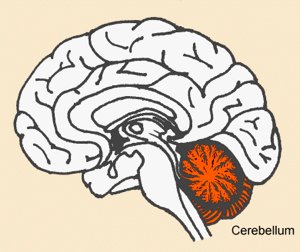
Primary Function
Coordinates movement by comparing intended actions with actual performance.
Regulates posture and balance.
Contributes to cognition and language processing.
Cerebrum
Composition: Hemispheres & Lobes
The cerebrum is divided into right and left hemispheres, separated by the longitudinal fissure. Each hemisphere contains lobes:
Frontal, Parietal, Temporal, Occipital, Insula
Surface Features
Cortex: Outer gray matter, highly folded.
Sulci: Shallow grooves (e.g., central sulcus).
Gyri: Raised folds (e.g., precentral and postcentral gyri).
Connections & Tracts
Corpus Callosum: Connects hemispheres.
Association tracts: Within same hemisphere.
Commissural tracts: Between hemispheres.
Projection tracts: Between cerebrum and lower CNS.
Basal Ganglia: Regulate movement initiation and termination; damage causes tremors (e.g., Parkinson’s Disease).
Functional Organization
Sensory Areas: Perception of somatosensory, visual, auditory, gustatory, and olfactory stimuli.
Motor Areas: Voluntary movement (primary motor area, Broca’s speech area).
Association Areas: Integration, memory, personality, reasoning, and complex learning.
Limbic System
Structure & Function
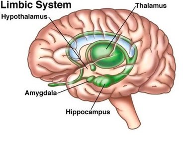
The limbic system is a network of structures involved in emotion, motivation, and memory.
Components: Cingulate gyrus, parahippocampal gyrus, dentate gyrus, amygdala, hippocampus, mammillary bodies, thalamic nuclei, olfactory bulbs, fornix.
Functions: Emotional responses (pain, pleasure, anger, affection); survival behaviors.
Diencephalon
Components & Functions
Thalamus: Major relay station for sensory impulses; involved in emotion, memory, and motor planning.
Hypothalamus: Controls autonomic functions, hormone production, emotional behavior, eating/drinking, body temperature, circadian rhythms.
Epithalamus: Contains pineal gland (melatonin secretion) and habenular nuclei (olfaction).
Circumventricular Organs (CVOs): Monitor blood chemistry; coordinate endocrine and nervous system homeostasis.
Cranial Nerves
General Information
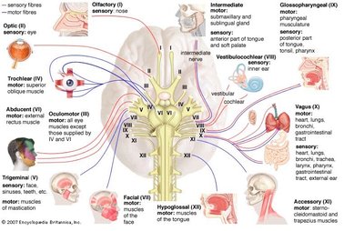
12 pairs; part of the peripheral nervous system.
Numbered from anterior to posterior.
Only CN I and II are purely sensory; others are mixed.
Sensory bodies in ganglia outside brain; motor bodies inside brain.
Major Cranial Nerves and Functions
Olfactory (I): Smell
Optic (II): Vision
Oculomotor (III): Eye movement, pupil constriction
Trochlear (IV): Eye movement
Trigeminal (V): Sensation from face, chewing
Abducens (VI): Eye movement
Facial (VII): Taste, facial expression
Vestibulocochlear (VIII): Hearing, equilibrium
Glossopharyngeal (IX): Taste, swallowing
Vagus (X): Taste, visceral sensation, heart rate, GI motility
Accessory (XI): Head and shoulder movement
Hypoglossal (XII): Tongue movement
Spinal Cord Anatomy
Protective Structures
Vertebral column: Bone and ligaments protect spinal cord.
Meninges: Cover spinal cord and nerve roots.
Epidural space: Fat cushion (not present around brain).
External Anatomy
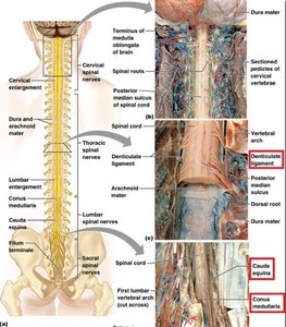
Cervical and lumbar enlargements: Nerves to upper and lower limbs.
Conus medullaris: Tapered end of spinal cord.
Filum terminale: Anchors spinal cord to coccyx.
Cauda equina: Nerve roots resembling a horse's tail.
Internal Anatomy
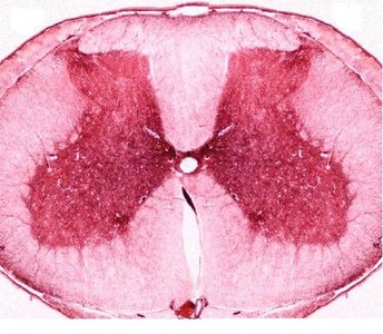
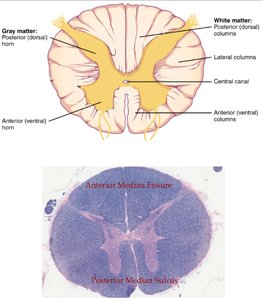
Gray matter: Central butterfly-shaped region; contains anterior and posterior horns, gray commissure.
White matter: Surrounds gray matter; divided into columns containing sensory (ascending) and motor (descending) tracts.
Central canal: Filled with CSF; continuous with fourth ventricle.
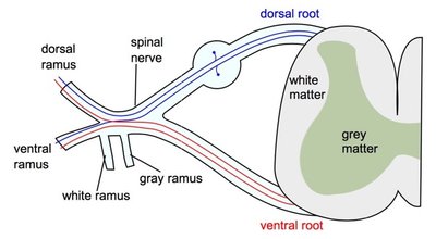
Spinal nerves: Posterior root (sensory), anterior root (motor); fusion forms mixed nerve.
Spinal Nerves
General Information
31 pairs: cervical, thoracic, lumbar, sacral, coccygeal.
Connect spinal cord to the rest of the body.
Roots: Dorsal (sensory, with ganglion), ventral (motor).
Coverings
Endoneurium: Around each axon.
Perineurium: Around each fascicle.
Epineurium: Around entire nerve.
Distribution: Branches and Plexuses
Rami: Posterior (dorsal) and anterior (ventral) branches.
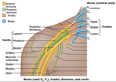
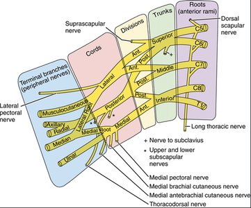
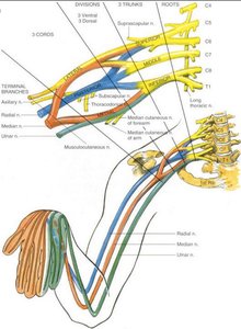
Plexuses: Cervical, brachial, lumbar, sacral, coccygeal; networks that recombine nerve fibers for specific body regions.
Reflexes & Reflex Arcs
Types of Reflex Arcs
Spinal, cranial, somatic, autonomic (visceral).
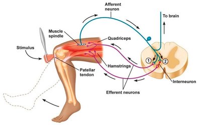
Basic Reflex Pathway
Sensory receptor
Sensory nerve
Integrating center (gray matter in CNS)
Motor nerve
Effector (skeletal muscle for somatic, smooth/cardiac/gland for autonomic)
Somatic Reflexes
Stretch reflex: Controls muscle length; monosynaptic, ipsilateral.
Tendon reflex: Controls muscle tension; polysynaptic, ipsilateral.
Flexor reflex: Withdrawal from pain; polysynaptic, intersegmental.
Crossed extensor reflex: Maintains balance; contralateral.
Diagnostic reflexes: Patellar, Achilles, Babinski, Abdominal.
Autonomic Reflexes
Regulate heart rate, GI motility, sphincter control, blood pressure, digestion, urination, defecation.
Pathway includes two motor nerves: preganglionic (CNS to ganglion), postganglionic (ganglion to effector).
Autonomic Nervous System (ANS)
Comparison: ANS vs Somatic Nervous System (SNS)
Feature | Somatic Nervous System | Autonomic Nervous System |
|---|---|---|
Sensory Input | Special & somatic senses (conscious) | Interoceptors (unconscious) |
Motor Pathway | Single, myelinated neuron to skeletal muscle | Two-neuron chain (preganglionic & postganglionic) to smooth/cardiac muscle, glands |
Effector | Skeletal muscle | Visceral organs |
Response | Always stimulates | Can stimulate or inhibit |
ANS Pathway Components
Preganglionic neuron: Cell body in CNS, axon to autonomic ganglion.
Postganglionic neuron: Cell body in ganglion, axon to effector.
Autonomic ganglia: Sympathetic (vertebral chain, collateral), parasympathetic (terminal/intramural).
Divisions of the ANS
Sympathetic: Fight or flight; thoracolumbar origin; wide distribution; ganglia distal to effector.
Parasympathetic: Rest and recovery; craniosacral origin; limited distribution; ganglia proximal to effector.
Cholinergic and Adrenergic Nerves & Receptors
Cholinergic: Release acetylcholine (ACh); receptors are nicotinic (excitation) and muscarinic (excitation/inhibition).
Adrenergic: Release norepinephrine (NE); receptors are alpha (a1, a2) and beta (B1, B2, B3); effects depend on receptor subtype.
Sensation and Perception
General Sensory Pathway
Stimulus → receptor → transduction → neural impulse → integration in cerebral cortex.
Association areas interpret and make sensory information useful.
Somesthetic Sensation
Receptors distributed throughout body; detect touch, pressure, stretch, movement, heat, cold, pain.
Unencapsulated (nociceptors, Merkel discs, hair receptors) and encapsulated (Meissner, Pacinian, Ruffini corpuscles).
Pathway: Receptors → spinal cord/brainstem → thalamus → postcentral gyrus.
Special Senses
Vision, hearing, taste, smell, equilibrium; originate in sense organs of the head.
Receptor Classification
By distribution: Somesthetic (general), special senses (head).
By location: Exteroceptors, interoceptors, visceroceptors, proprioceptors.
By modality: Mechanoreceptors, thermoreceptors, photoreceptors, chemoreceptors, osmoreceptors, nociceptors.
Homunculus
Somatotopy: Point-by-point correspondence between body and CNS.
Postcentral gyrus maps contralateral body regions.
