 뒤로
Chapter 11: Fundamentals of the Nervous System and Nervous Tissue
스터디 가이드 - 스마트 노트
 자료에 맞춘 맞춤형 노트, 핵심 정의, 예시, 맥락을 확장해 제공합니다.
The Nervous System: Overview
Functions of the Nervous System
The nervous system is responsible for coordinating the body's activities by transmitting signals between different parts of the body. It has three primary functions:
Sensory Input: Gathering information from sensory receptors about internal and external changes.
Integration: Processing and interpreting sensory input to determine an appropriate response.
Motor Output: Activating effector organs (muscles and glands) to produce a response.
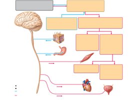
Divisions of the Nervous System
The nervous system is divided into two main parts:
Central Nervous System (CNS): Consists of the brain and spinal cord; serves as the integration and command center.
Peripheral Nervous System (PNS): Composed of paired spinal and cranial nerves that carry messages to and from the CNS.
Functional Divisions of the PNS
Sensory (Afferent) Division: Transmits impulses from sensory receptors to the CNS. Includes somatic afferent fibers (from skin, muscles, joints) and visceral afferent fibers (from organs).
Motor (Efferent) Division: Transmits impulses from the CNS to effector organs. Includes:
Somatic Nervous System: Voluntary control of skeletal muscles.
Autonomic Nervous System (ANS): Involuntary control of smooth muscle, cardiac muscle, and glands. Subdivided into sympathetic (mobilizes body systems during activity) and parasympathetic (conserves energy, promotes housekeeping functions during rest).
Histology of Nervous Tissue
Principal Cell Types
Neurons: Excitable cells that transmit electrical signals.
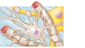
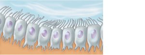
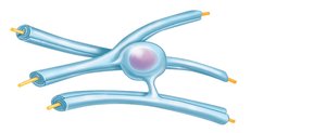
Neuroglia (Glial Cells): Supporting cells that provide structural and metabolic support for neurons. Types include:
Astrocytes (CNS): Most abundant; support neurons and maintain the blood-brain barrier.
Microglia (CNS): Defensive cells; act as macrophages.
Ependymal Cells (CNS): Line cerebrospinal fluid-filled cavities.
Oligodendrocytes (CNS): Form myelin sheaths around CNS nerve fibers.
Satellite Cells (PNS): Surround neuron cell bodies in the PNS.
Schwann Cells (PNS): Form myelin sheaths in the PNS.
Neurons: Structure and Function
Special Characteristics
Long-lived (can function for over 100 years)
Amitotic (do not divide after development, with few exceptions)
High metabolic rate (require continuous supply of oxygen and glucose)
Neuron Structure
Cell Body: Biosynthetic center and receptive region.
Dendrites: Short, branched processes; receptive regions that convey electrical signals toward the cell body as graded potentials.
Axon: Long process; conducting region that generates and transmits action potentials away from the cell body. Axon terminals are the secretory region, releasing neurotransmitters.
Bundles of Processes: Called tracts in the CNS and nerves in the PNS.
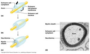
Myelin Sheath
The myelin sheath is a segmented protein-lipoid covering around most long or large-diameter axons, formed by Schwann cells in the PNS and oligodendrocytes in the CNS. Its functions include:
Protecting and electrically insulating the axon
Increasing the speed of nerve impulse transmission
Structural Classification of Neurons
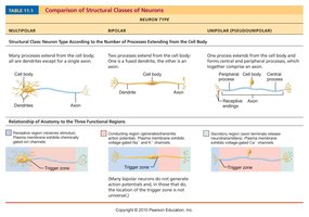
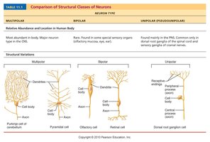
Neurons are classified based on the number of processes extending from the cell body:
Multipolar: One axon and several dendrites; most abundant; motor neurons and interneurons.
Bipolar: One axon and one dendrite; rare; found in retinal neurons.
Unipolar (Pseudounipolar): Single, short process with two branches; common in sensory neurons.
Functional Classification of Neurons
Sensory (Afferent): Transmit impulses from sensory receptors toward the CNS.
Motor (Efferent): Carry impulses from the CNS to effectors.
Interneurons (Association Neurons): Shuttle signals through CNS pathways; most are entirely within the CNS.
Membrane Potentials and Neural Signaling
Resting Membrane Potential
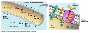
The resting membrane potential (Vr) is the potential difference across the membrane of a resting cell, typically –70 mV in neurons. It is generated by:
Differences in ion concentrations inside and outside the cell
Differential permeability of the plasma membrane
Changes in Membrane Potential
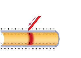
Depolarization: Reduction in membrane potential (toward zero); inside becomes less negative, increasing probability of producing a nerve impulse.
Hyperpolarization: Increase in membrane potential (away from zero); inside becomes more negative, reducing probability of producing a nerve impulse.
Graded Potentials
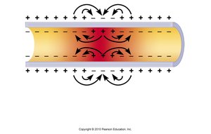
Graded potentials are short-lived, localized changes in membrane potential that spread as local currents change the membrane potential of adjacent regions. They can be depolarizations or hyperpolarizations.
Action Potentials
Action potentials (AP) are long-distance signals of axons that do not decrease in magnitude over distance. They are the principal means of neural communication.
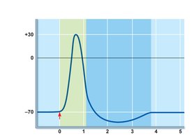
Resting Phase: Only leakage channels for Na+ and K+ are open; all gated channels are closed.
Depolarizing Phase: Local currents open Na+ channels; Na+ influx causes more depolarization.
Repolarizing Phase: Na+ channels close; K+ channels open, restoring internal negativity.
Hyperpolarization: Some K+ channels remain open, causing excessive K+ efflux.
All-or-None Response
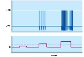
When a stimulus reaches threshold intensity, the impulse is full strength. More intense stimulation increases the rate of impulses, not their strength.
Refractory Period
Absolute Refractory Period: Membrane cannot be stimulated.
Relative Refractory Period: Membrane can be stimulated by a high-intensity stimulus.
Conduction Velocity
Axon Diameter: Larger diameter fibers conduct impulses faster.
Myelination: Myelinated axons conduct impulses faster than unmyelinated axons.
The Synapse
Structure and Function
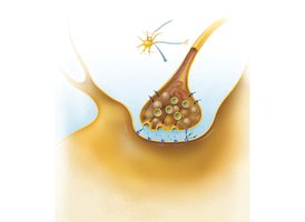
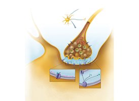
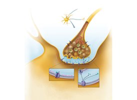
A synapse is a junction that mediates information transfer from one neuron to another or to an effector cell. It consists of:
Presynaptic Neuron: Conducts impulses toward the synapse.
Postsynaptic Neuron: Transmits impulses away from the synapse.
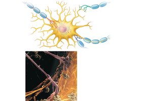
Chemical Synapses
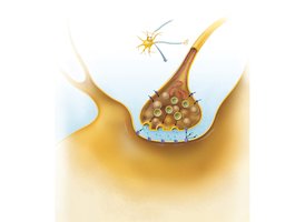
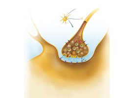
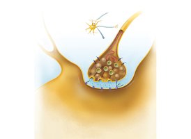
Chemical synapses are specialized for the release and reception of neurotransmitters. They include:
Axon terminal of the presynaptic neuron (contains synaptic vesicles)
Receptor region on the postsynaptic neuron
Synaptic cleft (fluid-filled space separating neurons)
Termination of Neurotransmitter Effects
Degradation by enzymes
Reuptake by astrocytes or axon terminal
Diffusion away from the synaptic cleft
Neurotransmitters
Chemical Classes
Acetylcholine (ACh): Released at neuromuscular junctions and some ANS neurons; degraded by acetylcholinesterase.
Biogenic Amines: Catecholamines (dopamine, norepinephrine, epinephrine) and indolamines (serotonin, histamine); involved in emotional behaviors and biological clock.
Amino Acids: GABA, glycine, aspartate, glutamate.
Peptides (Neuropeptides): Substance P (pain mediator), endorphins (natural opiates), gut-brain peptides (somatostatin, cholecystokinin).
Novel Messengers: ATP, nitric oxide (NO), carbon monoxide (CO).
Neural Integration
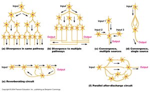
Functional Groups of Neurons
Neurons form functional groups that integrate incoming information and forward processed information to other destinations.
Homeostatic Imbalances
Multiple Sclerosis (MS)
An autoimmune disease affecting young adults
Symptoms: visual disturbances, weakness, loss of muscular control, speech disturbances, urinary incontinence
Myelin sheaths in the CNS become nonfunctional scleroses, leading to shunting and short-circuiting of nerve impulses
Impulse conduction slows and eventually ceases
