 뒤로
Chapter 11: Fundamentals of the Nervous System and Nervous Tissue - Study Notes
스터디 가이드 - 스마트 노트
 자료에 맞춘 맞춤형 노트, 핵심 정의, 예시, 맥락을 확장해 제공합니다.
The Nervous System: Overview
Introduction to the Nervous System
The nervous system is the primary controlling and communicating system of the body. It enables rapid and specific communication between cells via electrical and chemical signals, resulting in almost immediate responses. The nervous system generates nerve impulses (action potentials) to coordinate bodily functions.
Electrical signals: Action potentials transmitted along neurons.
Chemical signals: Neurotransmitters released at synapses.
Immediate responses: Enables quick reaction to stimuli.
Functions of the Nervous System
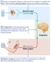
The nervous system performs three main functions: sensory input, integration, and motor output. These functions are tightly interconnected to maintain homeostasis and respond to environmental changes.
Sensory input: Gathering information from sensory receptors about internal and external changes.
Integration: Processing and interpretation of sensory input; decision-making occurs at the control center.
Motor output: Activation of effectors (muscles and glands) to produce a response.
Anatomical Organization of the Nervous System
Central and Peripheral Nervous Systems
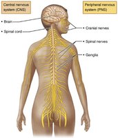
The nervous system is divided into two principal parts: the Central Nervous System (CNS) and the Peripheral Nervous System (PNS). Each has distinct anatomical structures and functions.
Central Nervous System (CNS): Composed of the brain and spinal cord, located in the dorsal body cavity. It serves as the integration and control center, interpreting sensory input and dictating motor output.
Peripheral Nervous System (PNS): Consists mainly of nerves extending from the brain and spinal cord. Includes spinal nerves (to/from spinal cord), cranial nerves (to/from brain), and the enteric nervous system (neurons in the gastrointestinal tract).
Functional Divisions of the Peripheral Nervous System
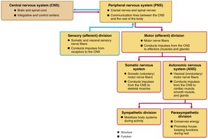
The PNS is further divided into sensory (afferent) and motor (efferent) divisions, each with specialized functions.
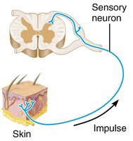
Sensory (afferent) division: Transmits impulses from sensory receptors to the CNS. Includes somatic sensory fibers (from skin, muscles, joints) and visceral sensory fibers (from internal organs).
Motor (efferent) division: Transmits impulses from the CNS to effectors (muscles and glands). Includes:
Somatic nervous system: Controls voluntary movements of skeletal muscles.
Autonomic nervous system: Controls involuntary functions of smooth muscle, cardiac muscle, and glands. Subdivided into sympathetic and parasympathetic divisions.
Cellular Structure of Nervous Tissue
Structure of a Motor Neuron
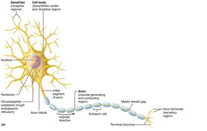
A neuron is an individual nerve cell, the basic structural and functional unit of the nervous system. Motor neurons have specialized regions for receiving, conducting, and transmitting signals.
Dendrites: Receptive regions that collect information.
Cell body (soma): Biosynthetic center and receptive region.
Axon: Conducting region that generates and transmits action potentials.
Axon terminals: Secretory region that releases neurotransmitters.
Neuroglia (Glial Cells)
Neuroglia are supporting cells in nervous tissue, essential for neuron function and health. They are classified based on their location and function.
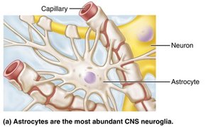
Astrocytes (CNS): Most abundant; support neurons, regulate exchanges with capillaries, control chemical environment, clean up potassium ions, and recycle neurotransmitters.
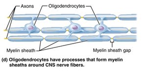
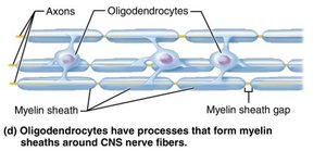
Oligodendrocytes (CNS): Form myelin sheaths around CNS axons, increasing conduction velocity.
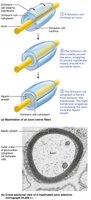
Schwann cells (PNS): Form myelin sheaths in the PNS, vital for regeneration of damaged nerve fibers.
Myelin Sheath and Neuron Processes
Myelin Sheath: Structure and Function
The myelin sheath is a white, fatty substance that coats many axons, especially long or large-diameter axons. It serves to protect and electrically insulate the axon, greatly increasing the speed of nerve impulse transmission.
Myelinated axons: Segmented sheath surrounds axons; rapid impulse conduction.
Nonmyelinated axons: Lack sheath; slower impulse conduction.
Myelin sheath gaps (nodes of Ranvier): Gaps between adjacent Schwann cells in PNS; sites for axon collateral emergence.
Myelination in the CNS
In the CNS, myelin is formed by oligodendrocyte processes, not whole cells. Each oligodendrocyte can wrap up to 60 axons at once. Thinnest fibers are unmyelinated but covered by extensions of neuroglia.
Neuron Structure and Functional Regions
Neuron Characteristics
Neurons are large, highly specialized cells that conduct impulses. They exhibit extreme longevity, high metabolic rates, and require continuous oxygen and glucose supply. All neurons have a cell body and one or more slender processes.
Cell body (soma): Contains nucleus and organelles; site of protein and neurotransmitter synthesis.
Dendrites: Short, branched processes; receptive regions for incoming signals.
Axon: Long process; conducting region for action potentials.
Axon terminals: Secretory region; releases neurotransmitters.
Functional Regions of a Neuron
Neurons have distinct functional regions:
Receptive region: Dendrites and cell body; receive signals.
Conducting region: Axon; transmits action potentials.
Secretory region: Axon terminals; release neurotransmitters.
Classification of Neurons
Structural Classification
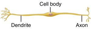
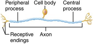
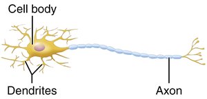
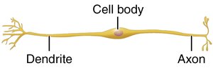
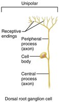
Neurons are classified based on the number of processes extending from the cell body:
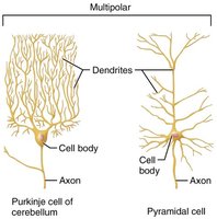
Multipolar neurons: One axon and two or more dendrites; most common type in CNS.
Bipolar neurons: One axon and one dendrite; found in special sensory organs.
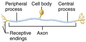
Unipolar neurons: One axon; cell body has no dendrites; most common type of sensory neuron.
Comparison of Structural Classes of Neurons
The following table summarizes the structural classes of neurons and their functional regions:
Neuron Type | Multipolar | Bipolar | Unipolar |
|---|---|---|---|
Processes | Many dendrites, one axon | One dendrite, one axon | One axon, no dendrites |
Location | CNS, motor neurons | Special sensory organs | Sensory neurons (PNS) |
Functional Regions | Receptive, conducting, secretory | Receptive, conducting, secretory | Receptive, conducting, secretory |
Functional Classification of Neurons
Sensory (Afferent) Neurons
Sensory neurons transmit impulses from sensory receptors toward the CNS. Almost all are unipolar neurons, specialized for rapid signal transmission from the periphery to the central nervous system.
Motor (Efferent) Neurons
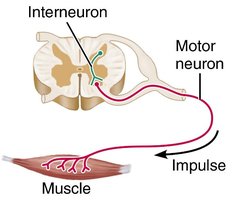
Motor neurons carry impulses from the CNS to effectors (muscles and glands). Most are multipolar neurons, responsible for activating muscle contraction or glandular secretion.
Interneurons
Interneurons (association neurons) are located within the CNS and connect sensory and motor neurons. They play a critical role in integration and processing of information.
Summary Table: Structural Classes of Neurons
Neuron Type | Multipolar | Bipolar | Unipolar |
|---|---|---|---|
Processes | Many dendrites, one axon | One dendrite, one axon | One axon, no dendrites |
Location | CNS, motor neurons | Special sensory organs | Sensory neurons (PNS) |
Functional Regions | Receptive, conducting, secretory | Receptive, conducting, secretory | Receptive, conducting, secretory |
Additional info: The notes above expand on the original content by providing definitions, examples, and context for each topic, ensuring completeness and academic quality for exam preparation.
