 뒤로
뒤로Chapter 14: Blood – Structure, Function, and Clinical Relevance
스터디 가이드 - 스마트 노트
 자료에 맞춘 맞춤형 노트, 핵심 정의, 예시, 맥락을 확장해 제공합니다.
Blood: Characteristics and Composition
Overview of Blood
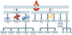
Blood is the only connective tissue with a liquid matrix, known as plasma. It plays a vital role in transporting substances, regulating homeostasis, maintaining interstitial fluid stability, and distributing heat throughout the body. The volume of blood varies based on body size, fluid and electrolyte concentrations, and adipose tissue content. In adults, blood constitutes about 8% of body weight, with typical volumes ranging from 4–5 liters in females and 5–6 liters in males.
Formed Elements: Blood cells produced in red bone marrow, including red blood cells (RBCs), white blood cells (WBCs), and platelets.
Plasma: The liquid matrix, making up about 55% of blood volume.
Components of Centrifuged Blood Samples
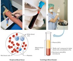
Blood samples are often centrifuged in clinical settings to detect abnormalities. The separation reveals distinct layers:
Plasma: Clear, straw-colored fluid at the top (about 55% of volume).
Red Blood Cells: Dense layer at the bottom (35–46% in females, 40–54% in males), known as the hematocrit (HCT).
Buffy Coat: Thin middle layer (<1% of volume) containing WBCs and platelets.
Formed Elements and Hematopoiesis
Origin and Differentiation of Blood Cells
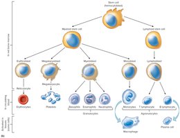
Blood cells originate from hematopoietic stem cells (hemocytoblasts) in red bone marrow. These stem cells differentiate into specialized cells under the influence of growth factors:
Lymphoid Stem Cells: Give rise to lymphocytes.
Myeloid Stem Cells: Produce RBCs, other WBCs, and platelets.
Red Blood Cells (Erythrocytes)
Structure and Function
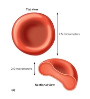
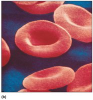
Red blood cells are biconcave discs, optimizing surface area for gas exchange. They lack nuclei and mitochondria, relying on glycolysis for ATP production. RBCs are primarily composed of hemoglobin, which binds and transports oxygen.
Oxyhemoglobin: Hemoglobin bound to O2.
Deoxyhemoglobin: Hemoglobin without O2.
Red Blood Cell Production and Regulation
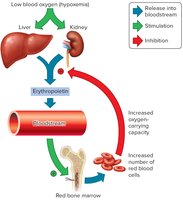
Erythropoiesis is the process of RBC formation, occurring in red bone marrow. Low blood oxygen triggers the release of erythropoietin (EPO) from the kidneys and liver, stimulating RBC production. This is regulated by a negative feedback mechanism.
Stages: Hemocytoblast → Erythroblast → Reticulocyte → Erythrocyte
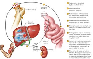
Life Span: Average of 120 days
Nutritional Requirements: Vitamin B12, folic acid (for DNA synthesis), and iron (for hemoglobin synthesis)
Anemia: Reduced O2-carrying capacity due to deficiency in RBCs or hemoglobin
Hemoglobin Breakdown and RBC Life Cycle
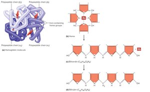
After their lifespan, RBCs are removed by the spleen or liver. Macrophages phagocytize worn-out cells, and hemoglobin is broken down into globin (amino acids) and heme (iron and biliverdin). Iron is recycled, and biliverdin is converted to bilirubin, which is excreted as bile pigment.
White Blood Cells (Leukocytes)
Types and Functions
White blood cells protect against disease and are produced in red bone marrow under hormonal control. They are categorized as granulocytes (neutrophils, eosinophils, basophils) and agranulocytes (lymphocytes, monocytes).
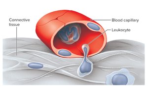
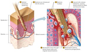
Diapedesis: WBCs squeeze through capillary walls to reach infection sites.
Phagocytosis: Neutrophils and monocytes engulf pathogens.
Inflammatory Response: Basophils release heparin and histamine to promote inflammation.
Chemotaxis: WBCs are attracted to infection sites by chemicals released from damaged cells.
Classification of White Blood Cells
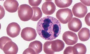
Neutrophils: First responders, strong phagocytes, 54–70% of WBCs
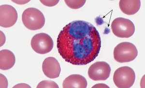
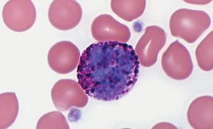
Eosinophils: Defend against parasitic worms, moderate allergic reactions, 1–3% of WBCs
Basophils: Release histamine and heparin, <1% of WBCs
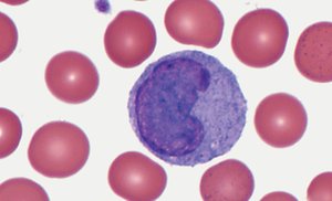
Monocytes: Largest WBCs, become macrophages, 3–9% of WBCs
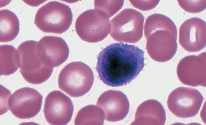
Lymphocytes: T cells (attack pathogens), B cells (produce antibodies), 25–33% of WBCs
Platelets (Thrombocytes)
Structure and Function
Platelets are cytoplasmic fragments of megakaryocytes, produced in response to thrombopoietin. They lack nuclei and are less than half the size of RBCs. Platelets are essential for hemostasis, adhering to broken surfaces and releasing serotonin to constrict blood vessels.
Normal Count: 150,000–400,000/µL
Thrombocytosis: High platelet count, risk of abnormal clot formation
Thrombocytopenia: Low platelet count, risk of easy bleeding and bruising
Blood Plasma
Composition and Functions
Plasma is the clear, straw-colored liquid portion of blood, comprising 55% of blood volume and 92% water. It transports nutrients, gases, hormones, and vitamins, and helps regulate fluid and electrolyte balance and maintain pH.
Plasma Proteins: Albumin, globulins, fibrinogen (not used as energy source)
Gases: Oxygen, carbon dioxide, nitrogen
Nutrients: Amino acids, simple sugars, nucleotides, lipids
Electrolytes: Sodium, potassium, calcium, magnesium, chloride, bicarbonate, phosphate, sulfate
Nonprotein Nitrogenous Substances: Urea, uric acid, amino acids, creatine, creatinine
Hemostasis
Mechanisms of Hemostasis
Hemostasis is the process of stopping bleeding, involving three main actions:
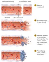
Vascular Spasm: Rapid contraction of smooth muscle in blood vessel walls after injury.
Platelet Plug Formation: Platelets adhere to rough surfaces and exposed collagen, forming a plug.
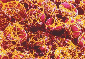
Blood Coagulation: Formation of a blood clot via a cascade of reactions, converting fibrinogen to fibrin.
Clotting Mechanisms
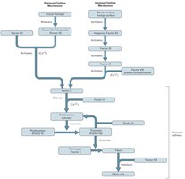
Extrinsic Mechanism: Triggered by tissue damage and release of tissue thromboplastin.
Intrinsic Mechanism: Initiated by contact with foreign surfaces, activating Hageman factor (XII).
Common Pathway: Both mechanisms converge to activate Factor X, leading to formation of fibrin mesh.
Blood Groups and Transfusions
ABO Blood Group
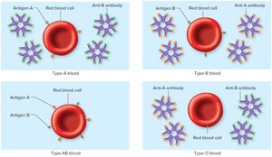
Blood types are determined by antigens (A and B) on RBC membranes. Safe transfusions require matching donor and recipient blood types to prevent agglutination.
Type A: Antigen A, anti-B antibodies
Type B: Antigen B, anti-A antibodies
Type AB: Antigens A and B, no antibodies (universal recipient)
Type O: No antigens, both anti-A and anti-B antibodies (universal donor)
Agglutination and Transfusion Reactions
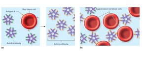
Agglutination occurs when antibodies in recipient plasma bind to donor RBC antigens, causing clumping and destruction of RBCs. Only ABO and Rh antigens cause serious transfusion reactions.
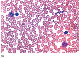
Rh Blood Group
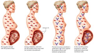
The Rh group is defined by the presence of antigen D. Rh-positive individuals have antigen D; Rh-negative individuals do not. Anti-Rh antibodies form only in Rh-negative people after exposure to Rh-positive blood, which can cause hemolytic disease of the newborn (erythroblastosis fetalis).
Summary Table: Blood Composition
Component | Percentage | Main Functions |
|---|---|---|
Plasma | 55% | Transport, regulation, fluid balance |
Red Blood Cells | 45% (Hematocrit) | Oxygen transport |
White Blood Cells | <1% | Immunity, defense |
Platelets | <1% | Hemostasis |
Key Equations
Hematocrit (HCT):
Blood Volume:
