 뒤로
뒤로Chapter 17: Blood – Structure, Function, and Disorders
스터디 가이드 - 스마트 노트
 자료에 맞춘 맞춤형 노트, 핵심 정의, 예시, 맥락을 확장해 제공합니다.
Blood: Composition and Functions
Overview of Blood Circulation
Blood is a vital fluid tissue responsible for transporting substances throughout the body. It circulates via arteries, capillaries, and veins, facilitating the exchange of oxygen, nutrients, and waste products. - Arteries carry oxygen-rich blood away from the heart. - Capillaries allow for exchange of gases and nutrients with tissues. - Veins return oxygen-deficient blood to the heart, which is then pumped to the lungs for gas exchange.
Components of Whole Blood
Blood consists of two main components: plasma and formed elements. - Plasma: The liquid portion, making up about 55% of blood volume. - Formed elements: Includes erythrocytes (RBCs), leukocytes (WBCs), and platelets. - Buffy coat: A thin layer containing leukocytes and platelets (<1% of blood). - Erythrocytes: Red blood cells, comprising about 45% of blood volume. 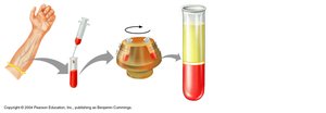
Physical Characteristics and Volume
Blood is a sticky, opaque fluid with a metallic taste. Its color varies with oxygen content: scarlet when oxygen-rich, dark red when oxygen-poor. - pH: 7.35–7.45 - Temperature: 38°C - Volume: 5–6 L in males, 4–5 L in females - Hematocrit: Percentage of blood volume occupied by RBCs (47% ± 5% in males, 42% ± 5% in females)
Functions of Blood
Blood serves three primary functions: 1. Distribution: Transports oxygen, nutrients, hormones, and waste products. 2. Regulation: Maintains body temperature, pH, and fluid balance. 3. Protection: Defends against infection and prevents blood loss via clotting.
Blood Plasma
Composition of Plasma
Plasma is 90% water and contains over 100 dissolved solutes, including nutrients, gases, hormones, wastes, proteins, and inorganic ions. - Plasma proteins are the most abundant solutes, produced mainly by the liver. - Albumin (60%): Maintains osmotic pressure and acts as a carrier. - Globulins (36%): Includes antibodies and transport proteins. - Fibrinogen (4%): Essential for blood clotting.
Formed Elements
Types of Formed Elements
The formed elements include erythrocytes, leukocytes, and platelets. - Erythrocytes (RBCs): Biconcave, anucleate cells specialized for oxygen transport. - Leukocytes (WBCs): Complete cells involved in immune defense. - Platelets: Cell fragments essential for clotting.
Erythrocytes (Red Blood Cells)
Erythrocytes are biconcave discs lacking nuclei and organelles, filled with hemoglobin for gas transport. Their shape increases surface area for efficient gas exchange. - Hemoglobin: Protein that binds oxygen and carbon dioxide. - Spectrin: Membrane protein providing flexibility. - ATP production: Anaerobic, so RBCs do not consume the oxygen they carry. 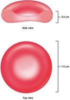
Erythrocyte Function and Hemoglobin
Hemoglobin consists of four polypeptide chains (two alpha, two beta), each with a heme group containing iron. - Each hemoglobin molecule can bind four oxygen molecules. - Oxyhemoglobin: Formed when oxygen binds in the lungs (ruby red). - Deoxyhemoglobin: Formed when oxygen is released in tissues (dark red). - Carbaminohemoglobin: Formed when carbon dioxide binds to hemoglobin.
Erythropoiesis: Red Blood Cell Production
Erythropoiesis is the process of RBC formation, occurring in red bone marrow. It involves several stages: 1. Hemocytoblast (stem cell) 2. Proerythroblast (committed cell) 3. Basophilic erythroblast (ribosome synthesis) 4. Polychromatic erythroblast (hemoglobin accumulation) 5. Orthochromatic erythroblast (ejection of nucleus) 6. Reticulocyte (immature RBC) 7. Erythrocyte (mature RBC) 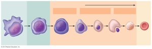
Regulation of Erythropoiesis
The balance between RBC production and destruction is regulated by hormones and nutrient availability. - Erythropoietin (EPO): Hormone released by kidneys in response to hypoxia (low oxygen). - Testosterone: Enhances EPO production, leading to higher RBC counts in males. - Dietary requirements: Iron, vitamin B12, folic acid, amino acids, lipids, and carbohydrates. 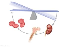
Life Cycle and Fate of Red Blood Cells
RBCs have a lifespan of 100–120 days. Old RBCs are removed by macrophages in the spleen. - Heme is degraded to bilirubin, which is excreted in bile. - Globin is broken down into amino acids. - Iron is salvaged and reused. 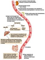
Erythrocyte Disorders
Anemia
Anemia is a condition of reduced oxygen-carrying capacity in blood, resulting in fatigue, pallor, and shortness of breath. - Causes: Blood loss, low RBC production, high RBC destruction. - Types: Hemorrhagic, iron-deficiency, pernicious, renal, aplastic, hemolytic, thalassemia, sickle-cell anemia.
Polycythemia
Polycythemia is an excess of RBCs, increasing blood viscosity. - Polycythemia vera: Bone marrow cancer. - Secondary polycythemia: Due to low oxygen (e.g., high altitude) or increased EPO. - Blood doping: Artificially increasing RBC count for athletic performance. 
Leukocytes (White Blood Cells)
Types and Functions
Leukocytes are complete cells involved in immune defense. They are classified as granulocytes or agranulocytes. - Granulocytes: Neutrophils, eosinophils, basophils. - Agranulocytes: Lymphocytes, monocytes. - Leukocytosis: Elevated WBC count, usually in response to infection. 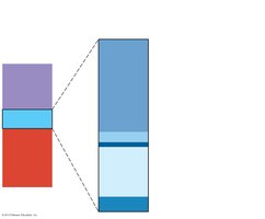
Summary of Formed Elements
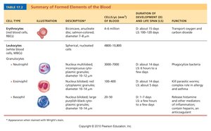
Cell Type | Description | Function |
|---|---|---|
Erythrocytes | Biconcave, anucleate | Transport O2 and CO2 |
Neutrophils | Multilobed nucleus, pale red granules | Phagocytize bacteria |
Eosinophils | Bilobed nucleus, red granules | Kill parasitic worms, modulate allergies |
Basophils | Lobed nucleus, blue granules | Release histamine, mediate inflammation |
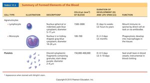
Cell Type | Description | Function |
|---|---|---|
Lymphocytes | Spherical nucleus, pale blue cytoplasm | Mount immune response |
Monocytes | U-shaped nucleus, gray-blue cytoplasm | Phagocytosis, activate lymphocytes |
Platelets | Disc-shaped fragments | Seal small tears, blood clotting |
Leukopoiesis: Formation of Leukocytes
Leukopoiesis is the process of WBC formation, stimulated by cytokines such as interleukins and colony-stimulating factors. - Hemocytoblasts differentiate into myeloid and lymphoid stem cells. - Myeloid stem cells produce granulocytes and monocytes. - Lymphoid stem cells produce lymphocytes. 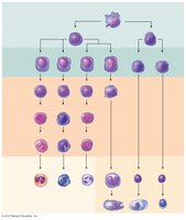
Platelets
Platelets are cytoplasmic fragments of megakaryocytes, essential for blood clotting. - Formation: Hemocytoblast → megakaryoblast → megakaryocyte → platelets. 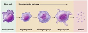
Hemostasis
Steps of Hemostasis
Hemostasis is the process of stopping bleeding, involving three steps: 1. Vascular spasm: Vasoconstriction of damaged vessel. 2. Platelet plug formation: Platelets adhere to exposed collagen and release chemicals. 3. Coagulation: Blood transforms from liquid to gel, forming a clot.
Coagulation Pathways
Coagulation involves intrinsic and extrinsic pathways, both leading to the formation of prothrombin activator. - Phase 1: Formation of prothrombin activator. - Phase 2: Conversion of prothrombin to thrombin. - Phase 3: Thrombin converts fibrinogen to fibrin, forming a mesh.
Clot Retraction and Repair
Clot retraction stabilizes the clot, and growth factors stimulate repair of the vessel wall.
Fibrinolysis
Fibrinolysis removes unneeded clots after healing. Plasminogen is converted to plasmin, which digests fibrin. 
Hemostasis Disorders
Thromboembolic Disorders
- Thrombus: Clot in an unbroken vessel. - Embolus: Thrombus that travels in the bloodstream. - Embolism: Embolus obstructs a vessel.
Bleeding Disorders
- Thrombocytopenia: Low platelet count. - Impaired liver function: Inability to produce clotting factors. - Hemophilia: Hereditary deficiency of clotting factors.
Disseminated Intravascular Coagulation (DIC)
DIC involves widespread clotting and severe bleeding, often as a complication of pregnancy, septicemia, or transfusions. 
Blood Transfusions and Blood Groups
Blood Transfusions
Whole-blood or packed RBC transfusions are used to restore oxygen-carrying capacity. Transfusion of incompatible blood can be fatal.
Human Blood Groups
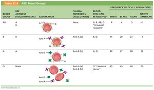
RBC membranes contain glycoprotein antigens (agglutinogens) used to classify blood into groups. - ABO blood groups: Determined by presence of A and B antigens. - Rh blood groups: Determined by presence of D antigen.
Blood Group | Antigens | Antibodies | Can Receive |
|---|---|---|---|
A | A | Anti-B | A, O |
B | B | Anti-A | B, O |
AB | A, B | None | A, B, AB, O |
O | None | Anti-A, Anti-B | O |
Hemolytic Disease of the Newborn
Occurs when an Rh– mother carries an Rh+ fetus. Anti-Rh antibodies can cross the placenta and attack fetal RBCs.
Transfusion Reactions
Mismatched blood transfusions cause agglutination and destruction of donor RBCs, leading to diminished oxygen capacity and possible renal failure.
Diagnostic Blood Tests
Common Blood Tests
- Hematocrit: Measures RBC percentage, used to diagnose anemia. - Blood glucose: Used for diabetes diagnosis. - Differential WBC count: Identifies types and numbers of WBCs. - Prothrombin time and platelet count: Assess hemostasis. - Complete blood count (CBC): Checks all formed elements, hematocrit, and hemoglobin. 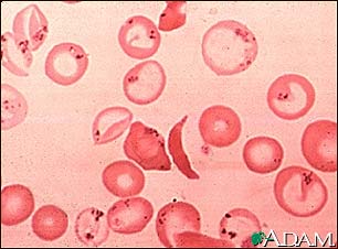
Developmental Aspects of Blood
Fetal and Aging Blood
- Before birth, blood cells form in the yolk sac, liver, and spleen. - By the seventh month, red bone marrow is the primary site. - Fetal hemoglobin (HbF) has higher oxygen affinity than adult hemoglobin. - Blood diseases of aging include chronic leukemias, anemias, and clotting disorders, often related to cardiovascular or immune system disorders.
