 뒤로
뒤로Chapter 20: The Lymphatic System and Immunity - Study Notes
스터디 가이드 - 스마트 노트
 자료에 맞춘 맞춤형 노트, 핵심 정의, 예시, 맥락을 확장해 제공합니다.
The Lymphatic System
Functions of the Lymphatic System
The lymphatic system is essential for maintaining fluid balance, absorbing fats, and supporting immune functions. It returns excess interstitial fluid to the bloodstream, absorbs lipid-soluble substances from the digestive tract, and filters pathogens from lymph and blood while housing and maturing immune cells.
Fluid Balance: Lymphatic vessels collect fluid from interstitial spaces and return it to circulation.
Fat Absorption: Specialized lymphatic capillaries called lacteals absorb fats from the digestive tract.
Immune Functions: Lymphatic tissues filter pathogens and support the maturation of lymphocytes.
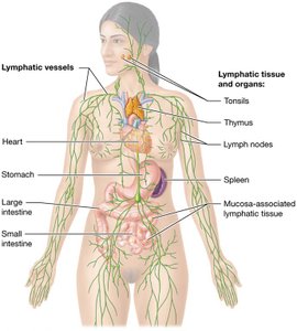
Anatomy of the Lymphatic System
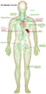
The lymphatic system consists of vessels, tissues, and organs such as tonsils, lymph nodes, spleen, and thymus. Lymph is the fluid transported by these vessels.
Lymphatic Vessels: Network that transports lymph.
Lymphatic Organs: Include tonsils, lymph nodes, spleen, and thymus.
Lymph: Fluid containing immune cells and solutes.
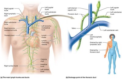
Lymphatic Vessels & Circulation
Lymphatic circulation begins in lymphatic capillaries, progresses through collecting vessels, trunks, and ducts, and ultimately drains into the venous system.
Sequence: Lymphatic capillaries → collecting vessels → trunks → ducts → subclavian veins.
Main Ducts: Thoracic duct and right lymphatic duct.
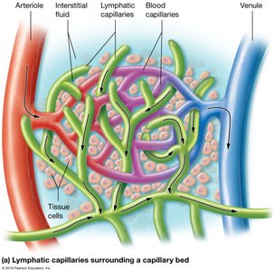
Lymphatic Capillaries
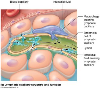
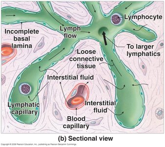
Lymphatic capillaries are highly permeable, blind-ended vessels found near blood capillaries. They allow one-way movement of fluid and are a low-pressure system without a central pump.
Structure: Overlapping endothelial cells form flaps that open and close based on pressure differences.
Permeability: Permit entry of cells, proteins, debris, pathogens, and immune cells.
Movement: Lymph moves via valves, skeletal muscle contraction, and smooth muscle activity.
Clinical Connection: Lymphedema
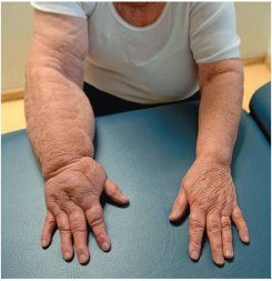
Lymphedema occurs when lymphatic vessels are removed or blocked, leading to accumulation of interstitial fluid and swelling in affected tissues.
Causes: Surgery, infection, or blockage.
Symptoms: Swelling, discomfort, and increased risk of infection.
Lymphoid Tissues and Organs
Structure and Function
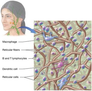
Lymphoid tissues are predominantly loose connective tissue containing lymphocytes, macrophages, dendritic cells, and reticular cells that produce reticular fibers.
Cell Types: B cells, T cells, macrophages, dendritic cells, reticular cells.
Function: Support immune cell development and pathogen filtration.

Mucosa-Associated Lymphatic Tissue (MALT)
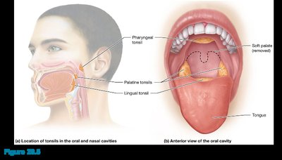
MALT consists of loosely organized clusters of lymphoid tissue that protect mucous membranes exposed to pathogens. Specialized MALT includes lymphoid follicles found in tonsils, Peyer's patches (ileum), and appendix.
Locations: Oral/nasal cavities, ileum, appendix.
Cell Types: Mainly B cells.
Lymph Nodes
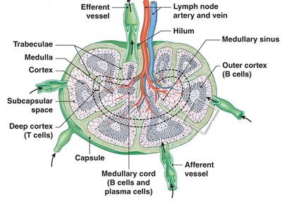
Lymph nodes are clusters of lymphatic tissue located along vessels. They filter lymph, trap pathogens, and house immune cells. Nodes can become swollen or painful during infection or cancer.
Locations: Axillary, cervical, inguinal, mesenteric regions.
Structure: Capsule, cortex (B cell-rich), medulla (macrophages, mature B cells).
Filtration: Lymph enters via afferent vessels, exits via efferent vessels.

Spleen
Structure and Function
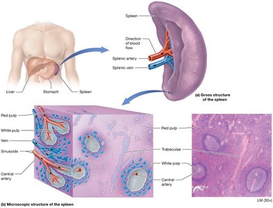
The spleen is the largest lymphoid organ, located in the left upper quadrant. It protects against blood pathogens, stores iron, initiates immune responses, and stores platelets.
Red Pulp: Contains macrophages to destroy old RBCs.
White Pulp: Filters blood pathogens and contains lymphocytes.
Thymus
Structure and Function
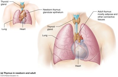
The thymus is a small, encapsulated organ in the mediastinum. It grows until puberty and then shrinks. It secretes thymosins, hormones that mature T cells.
Location: Mediastinum, above the heart.
Function: Maturation of T lymphocytes.

Immune System Overview
Types of Immunity
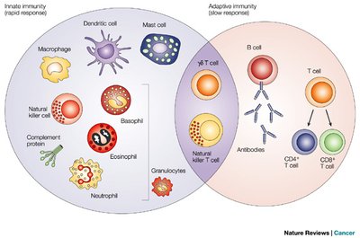
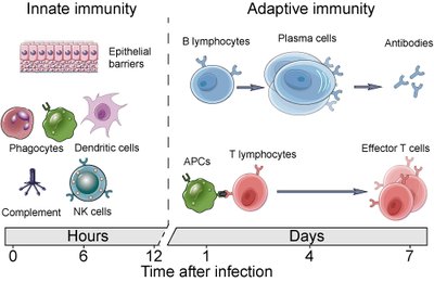
The immune system defends against trauma, pathogens, cancer cells, and toxins. Immunity is classified as innate (non-specific) or adaptive (specific/acquired).
Innate Immunity: Responds to all pathogens in the same way; includes surface barriers, cellular defenses, protein defenses, inflammation, and fever.
Adaptive Immunity: Responds to specific antigens; includes cell-mediated (T cells) and antibody-mediated (B cells) immunity.
Surface Barriers
Surface barriers are the first line of defense, including skin and mucous membranes. They provide mechanical and chemical protection against pathogens.
Skin: Resists mechanical stress; sebaceous glands secrete acidic substances.
Mucous Membranes: Trap pathogens; stomach mucosa is acidic; cilia move pathogens.
Cells and Proteins of the Immune System
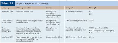
Immune cells include phagocytes (macrophages, neutrophils, eosinophils), dendritic cells, natural killer (NK) cells, and lymphocytes. Immune proteins include complement, antibodies, and cytokines.
Phagocytes: Ingest pathogens and debris.
NK Cells: Kill infected or abnormal cells.
Cytokines: Regulate immune cell activity.
Pathogens
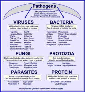
Pathogens are microscopic organisms that cause disease, including viruses, bacteria, fungi, protozoa, and multicellular parasites.
Viruses: Acellular, require host cells.
Bacteria: Prokaryotic cells.
Fungi: Eukaryotic, can infect tissues.
Protozoa: Single-celled eukaryotes.
Parasites: Multicellular organisms.
Innate Immunity (Non-specific Defenses)
Components and Mechanisms
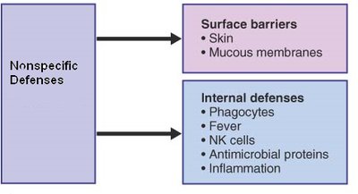
Innate immunity provides rapid, non-specific defense against pathogens. It includes surface barriers, cellular defenses, protein defenses, inflammation, and fever.
Surface Barriers: Skin and mucous membranes.
Cellular Defenses: Phagocytes, NK cells, mast cells, basophils, eosinophils.
Protein Defenses: Complement system, interferons.
Inflammatory Response: Local response to injury; four cardinal signs: redness, heat, swelling, pain.
Fever: Elevated body temperature due to pyrogens.
Phagocytosis and Cellular Defenses
Phagocytes ingest pathogens and debris. Neutrophils are most effective against bacteria, eosinophils against parasites. NK cells kill infected or abnormal cells.
Phagocytes: Macrophages, neutrophils, eosinophils.
NK Cells: Recognize and kill abnormal cells.
Protein Defenses: Complement and Interferons
The complement system consists of plasma proteins that, when activated, form a membrane attack complex (MAC) to lyse pathogens. Interferons are proteins released by infected cells to inhibit viral replication.
Complement: 30 proteins, activated in a cascade.
Interferons: Alpha, beta, gamma types; inhibit viral replication.
Inflammation and Fever
Inflammation is a local response to injury, characterized by redness, heat, swelling, and pain. Fever is a systemic response, raising body temperature to inhibit pathogens.
Inflammatory Mediators: Histamine, heparin, cytokines, prostaglandins.
Fever: Caused by pyrogens resetting hypothalamic temperature.
Adaptive Immunity (Specific Resistance)
Antigens and Properties
Adaptive immunity targets specific antigens and has four properties: specificity, versatility, memory, and tolerance.
Antigens: Substances recognized by B or T cells; can be peptides, polysaccharides, lipids, metals.
Self-antigens: Present on own cells, ignored by immune system.
Foreign-antigens: Generate immune response.
Properties: Specificity, versatility, memory, tolerance.
MHC Proteins
Major histocompatibility complex (MHC) proteins present antigens to T cells. Class I MHC is found on all nucleated cells; Class II MHC is found on antigen-presenting cells.
Class I MHC: Presents endogenous antigens; interacts with CD8+ T cells.
Class II MHC: Presents exogenous antigens; interacts with CD4+ T cells.
Cell-Mediated Immunity: T-Lymphocytes
T cells are divided into cytotoxic (CD8+) and helper (CD4+) types. Cytotoxic T cells destroy infected or abnormal cells; helper T cells stimulate other immune cells.
Cytotoxic T Cells: Release perforins, lymphotoxins, and induce apoptosis.
Helper T Cells: Secrete cytokines, activate B cells and other T cells.
Antibody-Mediated Immunity: B-Lymphocytes
B cells produce antibodies that bind to specific antigens. Antibodies consist of four polypeptide chains and have constant and variable regions.
Antibody Classes: IgG, IgM, IgA, IgE, IgD.
Functions: Neutralization, agglutination, opsonization, complement activation.
Immunological Memory
Memory B cells respond more efficiently upon re-exposure to the same antigen, forming the basis for vaccination.
Primary Response: First exposure, slower.
Secondary Response: Faster, stronger upon re-exposure.
Disorders of the Immune System
Hypersensitivity Disorders
Hypersensitivity disorders involve overreaction of the immune system, causing tissue damage. Types include immediate (Type I), antibody-mediated (Type II), immune complex (Type III), and delayed (Type IV) hypersensitivity.
Type I: Allergies, rapid response, IgE-mediated.
Type II: Antibody-mediated, slower onset.
Type III: Immune complex disorders, widespread inflammation.
Type IV: Delayed hypersensitivity, T cell-mediated.
Immunodeficiency Disorders
Immunodeficiency disorders result from decreased function of immune components. They can be primary (genetic) or secondary (acquired).
Primary: Hypogammaglobulinemia, SCID.
Secondary: HIV/AIDS, trauma, cancer, medications.
Autoimmune Disorders
Autoimmune disorders occur when the immune system attacks self-antigens, leading to tissue damage. Causes include exposure of sequestered antigens, molecular mimicry, inappropriate MHC expression, and nonspecific activation of B cells.
Stress and Aging Effects
Chronic stress and aging depress immune function, increasing vulnerability to infection and cancer. Glucocorticoids inhibit immune responses, and thymic hormone production decreases with age.
Effects: Reduced inflammatory response, fewer phagocytes, decreased antibody production.
