 뒤로
Chapter 21: The Immune System—Innate and Adaptive Body Defenses
스터디 가이드 - 스마트 노트
 자료에 맞춘 맞춤형 노트, 핵심 정의, 예시, 맥락을 확장해 제공합니다.
The Immune System: Overview
General Functions and Organization
The immune system is a functional system that provides resistance to disease-causing microorganisms, such as bacteria, fungi, and viruses. It is not an anatomical organ system, but rather a network of molecules and immune cells (especially lymphocytes) residing in lymphoid tissues and circulating in body fluids. The body employs three lines of defense, analogous to a medieval castle, to resist invasion: surface barriers, innate internal defenses, and adaptive defenses.
Surface Barriers: First line of defense, consisting of intact skin and mucosae.
Innate Internal Defenses: Second line, activated when surface barriers are breached.
Adaptive Defenses: Third line, highly specific and slower to respond.

Innate Defenses
Surface Barriers: First Line of Defense
Surface barriers include the skin and mucous membranes, along with their secretions. These barriers are present at birth and are highly effective at preventing entry of most microbes.
Keratinized Epidermis: Resistant to acids, bases, bacterial enzymes, and toxins.
Mucosae: Provide mechanical barriers within body tracts.
Protective Chemicals:
Acid Mantle: Acidity of skin, vaginal, and stomach secretions inhibits bacterial growth.
Enzymes: Lysozyme in saliva, mucus, and tears kills microbes; stomach enzymes digest pathogens.
Mucin: Sticky mucus traps microbes in digestive and respiratory tracts.
Defensins: Antimicrobial peptides secreted in response to barrier breach.
Other Chemicals: Sebum lipids and dermcidin in sweat are toxic to bacteria.
Respiratory Tract Modifications: Mucus-coated hairs in nose and cilia in upper tract trap and sweep out pathogens.
Innate Internal Defenses: Second Line of Defense
If surface barriers are breached, innate internal defenses are activated. These include phagocytes, natural killer cells, antimicrobial proteins, inflammation, and fever.
Pattern Recognition Receptors: Identify pathogens by recognizing specific molecular patterns (e.g., Toll-like receptors).
Phagocytes
Phagocytes are white blood cells that ingest and digest foreign invaders and debris.
Neutrophils: Most abundant, phagocytize infectious material.
Macrophages: Most voracious, derived from monocytes (free or fixed in tissues).
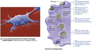
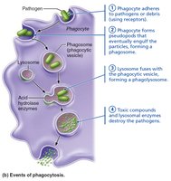
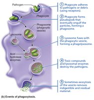
Phagocytosis Process
Phagocytosis involves several steps:
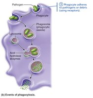
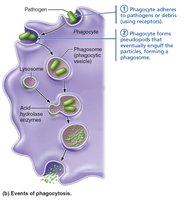
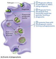
Phagocyte adheres to pathogen/debris.
Phagocyte forms pseudopods to engulf particles, forming a phagosome.
Phagosome fuses with lysosome, forming a phagolysosome.
Lysosomal enzymes and toxic compounds destroy pathogens.
Exocytosis removes indigestible material.
Opsonization
Some pathogens evade phagocytosis by hiding their surface markers. The immune system counters this by coating pathogens with opsonins (complement proteins or antibodies), which act as handles for phagocytes.
Natural Killer (NK) Cells
NK cells are large granular lymphocytes that police blood and lymph, killing cancer and virus-infected cells by inducing apoptosis and secreting chemicals to enhance inflammation.
Inflammation: Tissue Response to Injury
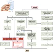
Inflammation is a nonspecific response to tissue injury, characterized by redness, heat, swelling, pain, and sometimes impaired function.
Benefits: Prevents spread of pathogens, disposes of debris, alerts adaptive immunity, and sets stage for repair.
Inflammatory Chemicals: Histamine, kinins, prostaglandins, cytokines, and complement.
Vasodilation and Permeability: Increased blood flow and capillary leakiness bring immune cells and proteins to the site.
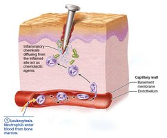
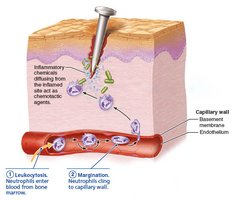
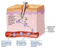
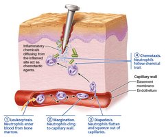
Phagocyte Mobilization: Steps include leukocytosis, margination, diapedesis, and chemotaxis.
Antimicrobial Proteins
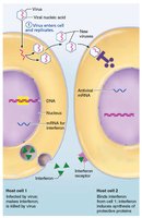
Interferons: Proteins produced by virus-infected cells that interfere with viral replication in neighboring cells and activate NK cells and macrophages.
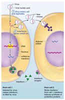
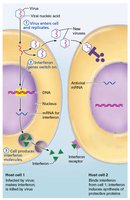
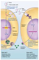
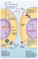
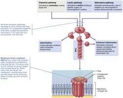
Complement System: A group of plasma proteins that, when activated, amplify inflammation, promote phagocytosis, and cause cell lysis. Activation occurs via classical, lectin, or alternative pathways, converging at C3.
Fever
Fever is a systemic response to infection, triggered by pyrogens released by leukocytes and macrophages. It increases metabolic rate, enhances immune response, and suppresses bacterial growth.
Adaptive Defenses
Key Features
The adaptive immune system is a specific defense system that targets and eliminates pathogens and abnormal cells. It consists of two overlapping arms:
Humoral Immunity: Antibody-mediated, targets extracellular pathogens.
Cellular Immunity: Cell-mediated, targets infected, cancerous, or foreign cells.
Antigens
Antigens are substances that trigger adaptive immune responses.
Complete Antigens: Have immunogenicity and reactivity; usually large, foreign proteins.
Haptens: Incomplete antigens that become immunogenic when attached to a protein carrier.
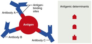
Antigenic Determinants: Specific parts of an antigen that antibodies or lymphocyte receptors bind to.
Self-Antigens: MHC Proteins
MHC proteins are glycoproteins on cell surfaces that display self or foreign peptides. They are unique to each individual and are essential for T cell recognition.
B and T Lymphocytes and Antigen-Presenting Cells
B Lymphocytes: Provide humoral immunity.
T Lymphocytes: Provide cellular immunity.
Antigen-Presenting Cells (APCs): Present antigens to T cells; include dendritic cells, macrophages, and B cells.
Lymphocyte Development, Maturation, and Activation
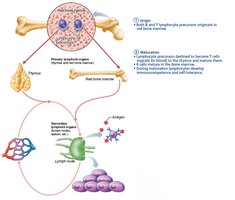
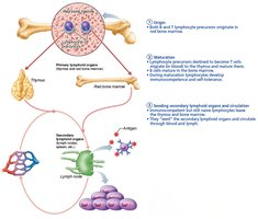
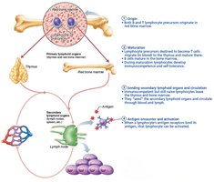
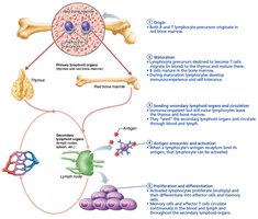
Lymphocytes undergo five general steps: origin, maturation, seeding secondary organs, antigen encounter and activation, and proliferation/differentiation. 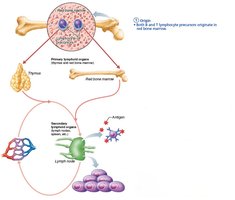
T Cell Education in the Thymus
T cells undergo positive and negative selection to ensure immunocompetence and self-tolerance. 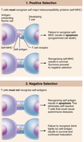
Humoral Immunity
Activation and Differentiation of B Cells
When a B cell encounters its antigen, it undergoes clonal selection, proliferates, and differentiates into plasma cells (antibody-secreting) and memory cells. 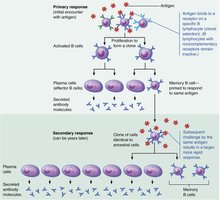
Immunological Memory
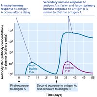
Primary Response: Slow, occurs upon first exposure to antigen.
Secondary Response: Faster and stronger, due to memory cells.
Active and Passive Humoral Immunity
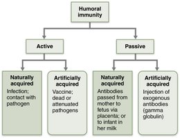
Active Immunity: Body produces antibodies (natural via infection, artificial via vaccination).
Passive Immunity: Antibodies are introduced (natural via placenta/milk, artificial via injection).
Antibodies (Immunoglobulins)
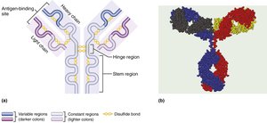
Antibodies are proteins secreted by plasma cells, grouped into five classes: IgG, IgA, IgM, IgE, and IgD.
Structure: Four polypeptide chains (two heavy, two light), forming a Y-shaped molecule with antigen-binding sites.
Functions: Neutralization, agglutination, precipitation, complement activation.
Cellular Immunity
T Lymphocytes
T cells provoke cellular immune responses, directly killing infected, cancerous, or foreign cells, or regulating immune responses.
CD4 Cells: Become helper T cells or regulatory T cells.
CD8 Cells: Become cytotoxic T cells.
MHC Proteins and Antigen Presentation
T cells respond only to antigens presented on MHC proteins.
MHC Class I: Present endogenous antigens to CD8 cells.
MHC Class II: Present exogenous antigens to CD4 cells.
Activation and Differentiation of T Cells
T cell activation requires antigen binding and co-stimulation, leading to proliferation and differentiation into effector and memory cells.
Roles of Specific Effector T Cells
Helper T Cells: Mobilize both humoral and cellular immunity, activate B and T cells, and secrete cytokines.
Cytotoxic T Cells: Directly attack and kill infected and cancerous cells using perforins and granzymes.
Regulatory T Cells: Suppress immune responses, prevent autoimmunity.
Immune System Disorders
Immunodeficiencies
SCID: Severe combined immunodeficiency, marked deficit in B and T cells.
AIDS: Acquired immune deficiency syndrome, caused by HIV, destroys helper T cells.
Autoimmune Diseases
Immune system fails to distinguish self from foreign, producing autoantibodies and self-reactive T cells. Examples include rheumatoid arthritis, type 1 diabetes, and lupus.
Hypersensitivities
Immediate (Acute) Hypersensitivity: Allergies, mediated by IgE and histamine release.
Subacute Hypersensitivity: Mediated by IgM and IgG, slower onset.
Delayed Hypersensitivity: Mediated by T cells, takes 1–3 days to appear.
Developmental Aspects
Immune System Development and Aging
Stem cells develop in liver and spleen early, then bone marrow.
Immune efficiency wanes with age, increasing susceptibility to infection, cancer, and autoimmune diseases.
Additional info: Academic context was added to clarify mechanisms, definitions, and clinical relevance. Tables referenced in the original file were summarized in text, as their main content was incorporated into explanations.
