 뒤로
Chapter 22: The Respiratory System – Anatomy & Physiology Study Guide
스터디 가이드 - 스마트 노트
 자료에 맞춘 맞춤형 노트, 핵심 정의, 예시, 맥락을 확장해 제공합니다.
The Respiratory System: Overview
Major Functions and Processes
The respiratory system is essential for gas exchange, supplying oxygen to cells for cellular respiration and removing carbon dioxide, a metabolic waste. It works closely with the cardiovascular system to ensure efficient transport and exchange of gases throughout the body.
Pulmonary ventilation: Movement of air into and out of the lungs (breathing).
Pulmonary gas exchange: Exchange of gases (O2 and CO2) between lungs and blood.
Transport of respiratory gases: Movement of O2 and CO2 in blood via the cardiovascular system.
Tissue gas exchange: Exchange of gases between blood and tissues.
Functional Anatomy of the Respiratory System
Major Organs and Divisions
The respiratory system is divided into upper and lower regions, each with specialized structures for air conduction, filtration, and gas exchange.
Upper respiratory system: Nose, paranasal sinuses, pharynx
Lower respiratory system: Larynx, trachea, bronchi, lungs, alveoli
Respiratory muscles: Classified as part of the muscular system, essential for ventilation
Upper Respiratory System
The Nose and Paranasal Sinuses
The nose is the only externally visible part of the respiratory system and serves multiple functions, including air conduction, filtration, and olfaction.
Functions: Provides airway, moistens and warms air, filters and cleans inspired air, serves as a resonating chamber for speech, houses olfactory receptors.
Regions: External nose and nasal cavity
Clinical Note: Rhinitis and Sinusitis
Rhinitis: Inflammation of nasal mucosa, leading to excessive mucus, congestion, and postnasal drip.
Sinusitis: Inflammation of paranasal sinuses, often secondary to rhinitis; can cause sinus headaches due to blocked air pressure.
The Pharynx
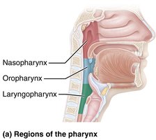
The pharynx is a muscular tube that connects the nasal cavity and mouth to the larynx and esophagus. It is divided into three regions: nasopharynx, oropharynx, and laryngopharynx.
Nasopharynx: Posterior to nasal cavity; passage for air only.
Oropharynx: Posterior to oral cavity; passage for air and food.
Laryngopharynx: Posterior to larynx; passage for air and food.
Clinical Note: Adenoids
Enlarged adenoids: Can block air passage, forcing mouth breathing and affecting speech and sleep.
Lower Respiratory System
Conducting and Respiratory Zones
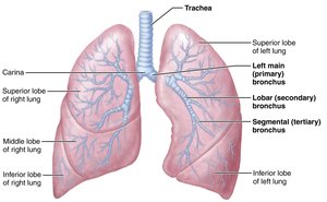
The lower respiratory system is divided anatomically and functionally into conducting and respiratory zones.
Conducting zone: All airways from nose to respiratory bronchioles; transports, cleanses, warms, and humidifies air.
Respiratory zone: Sites of gas exchange; includes respiratory bronchioles, alveolar ducts, and alveoli.
The Larynx
The larynx, or voice box, is responsible for maintaining an open airway, routing air and food, and voice production.
Structure: Extends from the hyoid bone to the trachea; opens into the laryngopharynx.
Functions: Airway patency, routing air/food, voice production (vocal folds).
Movements of the Vocal Folds
Vocal folds vibrate to produce sound; pitch and loudness are determined by their tension and the force of airflow.
Pitch: Determined by length and tension of vocal folds.
Loudness: Depends on force of airflow.
Resonating chambers: Pharynx, oral, nasal, and sinus cavities amplify sound.
Clinical Note: Laryngitis
Laryngitis: Inflammation of vocal folds, causing hoarseness or loss of voice; often due to infection or overuse.
Tissue Composition of the Tracheal Wall
The trachea is supported by cartilaginous rings and lined with ciliated epithelium to trap and remove debris.
Layers: Mucosa, submucosa, hyaline cartilage, adventitia.
Cilia: Move mucus upward toward the throat.
Clinical Note: Smoking and Tracheal Obstruction
Smoking: Destroys cilia, leading to mucus accumulation and "smoker's cough."
Tracheal obstruction: Life-threatening; Heimlich maneuver can expel obstructions.
Conducting Zone Passages
Respiratory Zone Structures
The respiratory zone includes alveoli, the primary site of gas exchange. Alveoli are surrounded by capillaries and elastic fibers, and contain pores for pressure equalization.
Respiratory membrane: Thin barrier for gas exchange; consists of alveolar and capillary walls.
Type I alveolar cells: Simple squamous epithelium for gas exchange.
Type II alveolar cells: Secrete surfactant and antimicrobial proteins.
Alveolar macrophages: Remove debris and pathogens.
The Pleurae
Pleural Structure and Function
The pleurae are serous membranes that surround the lungs and line the thoracic cavity, providing lubrication and surface tension to facilitate breathing.
Parietal pleura: Lines thoracic wall and diaphragm.
Visceral pleura: Covers external lung surface.
Pleural fluid: Lubricates and prevents separation of pleurae.
Clinical Note: Pleurisy and Pleural Effusion
Pleurisy: Inflammation of pleurae, causing pain and friction.
Pleural effusion: Accumulation of fluid in pleural cavity, hindering breathing.
Respiratory Physiology
Pulmonary Ventilation: Mechanics of Breathing
Pulmonary ventilation consists of inspiration and expiration, driven by changes in thoracic volume and pressure.
Inspiration: Active process; diaphragm and external intercostals contract, increasing thoracic volume.
Expiration: Passive during quiet breathing; muscles relax, thoracic volume decreases.
Forced expiration: Active; uses abdominal and internal intercostal muscles.
Pressure Relationships in the Thoracic Cavity
Intrapulmonary pressure (Ppul): Pressure in alveoli; equalizes with atmospheric pressure.
Intrapleural pressure (Pip): Pressure in pleural cavity; always negative relative to Ppul.
Transpulmonary pressure: Difference between Ppul and Pip; keeps lungs inflated.
Pneumothorax and Atelectasis
Pneumothorax: Air in pleural cavity causes lung collapse; treated by removing air.
Atelectasis: Collapse of lung or alveoli due to obstruction or pneumothorax.
Physical Factors Influencing Pulmonary Ventilation
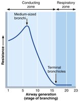
Airway resistance: Usually insignificant; greatest in medium-sized bronchi.
Alveolar surface tension: Surfactant reduces surface tension, preventing alveolar collapse.
Lung compliance: Measure of lung expandability; decreased by fibrosis, surfactant deficiency, or thoracic deformities.
Pulmonary Volumes and Capacities
Measuring Ventilation
Pulmonary volumes and capacities are measured to assess respiratory health. Spirometry is used to evaluate these parameters.
Tidal volume (TV): Air moved in/out per breath (~500 mL).
Inspiratory reserve volume (IRV): Air forcibly inspired beyond TV (2100–3200 mL).
Expiratory reserve volume (ERV): Air forcibly expired beyond TV (1000–1200 mL).
Residual volume (RV): Air remaining in lungs after forced expiration.
Pulmonary Volume | Adult Male Average | Adult Female Average | Description |
|---|---|---|---|
Tidal Volume (TV) | 500 mL | 500 mL | Amount of air inhaled or exhaled with each breath under resting conditions |
Inspiratory Reserve Volume (IRV) | 3100 mL | 1900 mL | Amount of air that can be forcefully inhaled after a normal tidal volume inhalation |
Expiratory Reserve Volume (ERV) | 1200 mL | 700 mL | Amount of air that can be forcefully exhaled after a normal tidal volume exhalation |
Residual Volume (RV) | 1200 mL | 1100 mL | Amount of air remaining in the lungs after a forced exhalation |
Pulmonary Capacities
Inspiratory capacity (IC): TV + IRV
Functional residual capacity (FRC): RV + ERV
Vital capacity (VC): TV + IRV + ERV
Total lung capacity (TLC): TV + IRV + ERV + RV
Dead Space
Anatomical dead space: Conducting zone airways; no gas exchange.
Alveolar dead space: Nonfunctional alveoli; can increase with disease.
Total dead space: Sum of anatomical and alveolar dead space.
Gas Exchange: Diffusion Between Blood, Lungs, and Tissues
Basic Properties of Gases
Dalton’s law: Total pressure of a gas mixture equals the sum of partial pressures of individual gases.
Henry’s law: Gas dissolves in liquid in proportion to its partial pressure and solubility.
Partial Pressure Gradients
Oxygen: Steep gradient drives diffusion into blood.
Carbon dioxide: Smaller gradient, but CO2 is more soluble, so diffuses efficiently.
Clinical Note: Emphysema and Surface Area
Emphysema: Destruction of alveolar walls reduces surface area, impairing gas exchange.
Ventilation-Perfusion Coupling
Optimal gas exchange: Requires matching ventilation (air flow) and perfusion (blood flow).
Local autoregulation: Adjusts arteriolar and bronchiolar diameter based on O2 and CO2 levels.
Oxygen and Carbon Dioxide Transport
Oxygen Transport
Oxygen is transported in blood primarily bound to hemoglobin (Hb) in red blood cells.
98.5% bound to Hb; 1.5% dissolved in plasma.
Oxyhemoglobin: Hb bound to O2.
Deoxyhemoglobin: Hb not bound to O2.
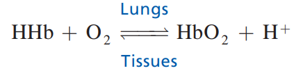
Equation for oxygen binding:
Oxygen-Hemoglobin Dissociation Curve
The relationship between partial pressure of oxygen and hemoglobin saturation is sigmoidal, reflecting cooperative binding.
Arterial blood: Hb is 98% saturated.
Venous blood: Hb is 75% saturated (venous reserve).
Factors affecting curve: Temperature, pH, CO2, and BPG.
Carbon Dioxide Transport
Transported in three ways: Dissolved in plasma, bound to Hb, as bicarbonate ion (HCO3-).
Bicarbonate buffer system: Maintains blood pH.
Clinical Conditions and Respiratory Diseases
Hypoxia
Definition: Inadequate oxygen delivery to tissues.
Types: Anemic, ischemic, histotoxic, hypoxemic.
Carbon monoxide poisoning: Hb binds CO with higher affinity than O2.
Chronic Obstructive Pulmonary Disease (COPD)
Emphysema: Alveolar destruction, reduced elasticity, hyperinflation.
Chronic bronchitis: Excess mucus, inflammation, obstructed airways.
Symptoms: Dyspnea, hypoventilation, respiratory acidosis, hypoxemia.
Treatment: Bronchodilators, corticosteroids, oxygen therapy.
Asthma
Characteristics: Reversible airway obstruction, inflammation, bronchospasms.
Treatment: Inhaled corticosteroids, bronchodilators.
Tuberculosis (TB)
Cause: Mycobacterium tuberculosis infection.
Symptoms: Fever, cough, weight loss, hemoptysis.
Lung Cancer
Types: Adenocarcinoma, squamous cell carcinoma, small cell carcinoma.
Risk factors: Smoking, environmental pollutants.
Treatment: Surgery, radiation, chemotherapy, targeted drug therapies.
Sleep Apnea
Obstructive sleep apnea: Collapse of upper airway during sleep.
Central sleep apnea: Reduced drive from brain stem respiratory centers.
Treatment: CPAP device.
Summary Table: Pulmonary Volumes and Capacities
Capacity | Formula | Description |
|---|---|---|
Inspiratory Capacity (IC) | TV + IRV | Amount of air that can be inspired after normal tidal expiration |
Functional Residual Capacity (FRC) | RV + ERV | Amount of air remaining in lungs after normal tidal expiration |
Vital Capacity (VC) | TV + IRV + ERV | Total amount of exchangeable air |
Total Lung Capacity (TLC) | TV + IRV + ERV + RV | Sum of all lung volumes |
Key Equations
Dalton’s Law:
Henry’s Law: (where C = concentration, k = solubility constant, P = partial pressure)
Lung Compliance:
Additional info:
Some clinical notes and disease mechanisms were expanded for clarity and completeness.
Tables were recreated and summarized based on textbook context.
