 뒤로
Chapter 23: The Digestive System – Anatomy & Physiology Study Guide
스터디 가이드 - 스마트 노트
 자료에 맞춘 맞춤형 노트, 핵심 정의, 예시, 맥락을 확장해 제공합니다.
The Digestive System
Anatomy of the Gastrointestinal (GI) Tract
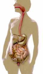
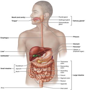
The digestive system consists of the gastrointestinal (GI) tract, also known as the alimentary canal, and accessory organs. The GI tract is a continuous tube extending from the mouth to the anus, responsible for the digestion and absorption of food.
Organs of the GI tract: Mouth, pharynx, esophagus, stomach, small intestine, large intestine, rectum, anus
Accessory structures: Teeth, tongue, gallbladder, salivary glands, liver, pancreas
GI Tract Activities
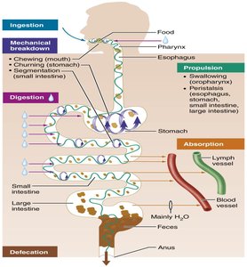
The digestive process involves several coordinated activities that ensure the breakdown and absorption of nutrients.
Ingestion: Taking food into the mouth
Mechanical breakdown: Chewing (mouth), churning (stomach), segmentation (small intestine)
Propulsion: Swallowing (pharynx), peristalsis (esophagus, stomach, intestines)
Digestion: Enzymatic breakdown of food
Absorption: Movement of nutrients into blood or lymph
Defecation: Elimination of indigestible substances
Peristalsis and Segmentation
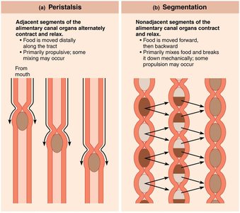
Two main types of muscular contractions move and mix food along the GI tract:
Peristalsis: Adjacent segments of the alimentary canal organs contract and relax, propelling food distally.
Segmentation: Nonadjacent segments contract and relax, mixing food and breaking it down mechanically.
Histology of the Alimentary Canal
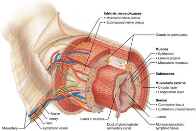
The wall of the alimentary canal consists of four basic layers: mucosa, submucosa, muscularis externa, and serosa. Each layer has specialized functions for digestion and absorption.
Mucosa: Epithelium, lamina propria, muscularis mucosae
Submucosa: Connective tissue, glands, blood vessels
Muscularis externa: Circular and longitudinal muscle layers
Serosa: Outermost layer (mesothelium)
Intrinsic nerve plexuses: Myenteric and submucosal plexuses regulate GI motility
Neural Reflex Pathways in the GI Tract
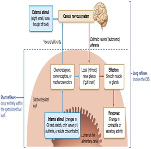
Digestive activities are regulated by neural reflexes initiated by stimuli inside or outside the GI tract. These involve both short (local) and long (central) reflexes.
External stimuli: Sight, smell, taste, thought of food
Internal stimuli: GI tract stretch, changes in lumen pH, nutrients
Effectors: Smooth muscle or glands
Response: Changes in contractile or secretory activity
The Mouth and Associated Structures
Anatomy of the Oral Cavity
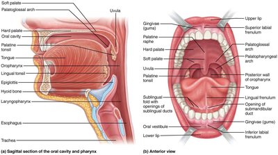
The mouth is the entry point for food and is involved in mechanical and chemical digestion. It contains the teeth, tongue, and salivary glands.
Structures: Hard and soft palate, uvula, gingivae (gums), tongue, tonsils
Functions: Mastication (chewing), mixing food with saliva, swallowing
Tongue and Papillae
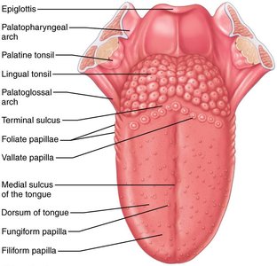
The tongue is a muscular organ that aids in food manipulation, taste, and swallowing. It contains various types of papillae, some of which house taste buds.
Types of papillae: Filiform (do not contain taste buds), fungiform, vallate, foliate
Functions: Taste, mechanical manipulation, speech
Salivary Glands
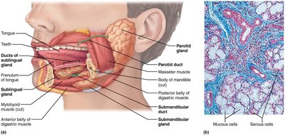
Salivary glands produce saliva, which contains enzymes, antimicrobial agents, and mucus. Saliva begins the chemical digestion of carbohydrates and lubricates food for swallowing.
Major glands: Parotid, submandibular, sublingual
Saliva: Contains amylase (digests starch), lysozyme, IgA, mucins
Xerostomia: Dry mouth due to reduced saliva production
Teeth and Dentition
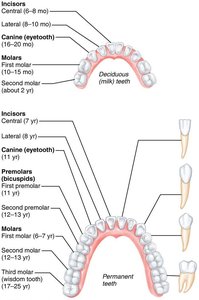
Teeth are essential for mechanical digestion. Humans have two sets of teeth: deciduous (milk) teeth and permanent teeth.
Deciduous teeth: 20 teeth, erupt in childhood
Permanent teeth: 32 teeth, replace deciduous teeth
Types: Incisors, canines, premolars, molars
Tooth Structure
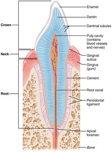
Each tooth consists of a crown, neck, and root. The crown is covered by enamel, the hardest substance in the body.
Enamel: Hard, outer layer
Dentin: Underlies enamel, forms bulk of tooth
Pulp cavity: Contains blood vessels and nerves
Cement: Covers root, attaches tooth to periodontal ligament
Tooth and Gum Disease
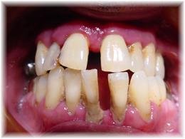
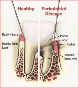
Dental diseases include cavities, plaque, calculus, gingivitis, and periodontal disease. These conditions can affect overall health and increase the risk of systemic diseases.
Cavities: Decay of tooth structure
Plaque: Sticky film of bacteria
Calculus: Hardened plaque (tartar)
Gingivitis: Inflammation of gums
Periodontal disease: Advanced gum disease, affects bone and ligaments
Esophagus and Swallowing
Microscopic Anatomy of the Esophagus
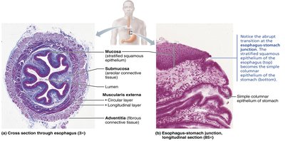
The esophagus is a muscular tube that transports food from the pharynx to the stomach. Its wall consists of mucosa, submucosa, muscularis externa, and adventitia.
Mucosa: Stratified squamous epithelium
Transition: Abrupt change to simple columnar epithelium at the stomach
Deglutition (Swallowing)
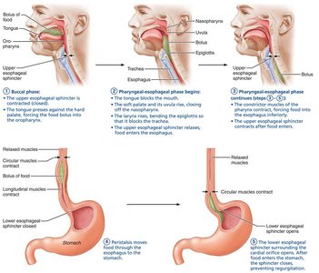
Swallowing is a complex process involving the mouth, pharynx, and esophagus. It consists of voluntary and involuntary phases.
Buccal phase: Voluntary, food is pushed into the pharynx
Pharyngeal-esophageal phase: Involuntary, food moves through pharynx and esophagus
Sphincters: Upper and lower esophageal sphincters regulate passage
Stomach
Gross Anatomy of the Stomach
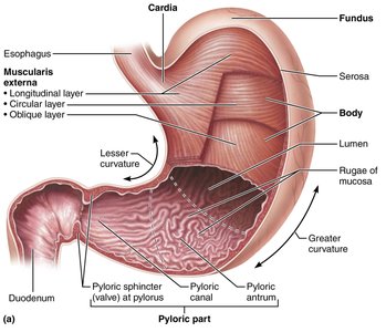
The stomach is a muscular organ that stores, mixes, and digests food. It has several regions and specialized structures.
Regions: Cardia, fundus, body, pyloric part
Rugae: Folds in the mucosa that allow expansion
Sphincters: Cardiac (gastroesophageal) and pyloric sphincters
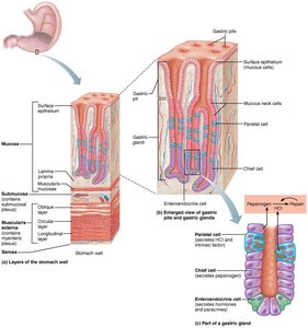
Microscopic Anatomy of the Stomach
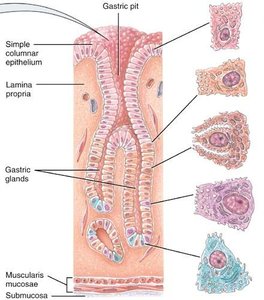
The stomach wall contains specialized cells that secrete various substances for digestion.
Surface mucous cells: Secrete thick alkaline mucus
Mucous neck cells: Secrete thin acidic mucus
Parietal cells: Secrete hydrochloric acid (HCl) and intrinsic factor
Chief cells: Secrete pepsinogen and lipases
Enteroendocrine cells: Secrete hormones (gastrin, histamine, serotonin)
Gastritis and Gastric Ulcers
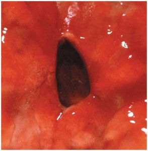
Gastritis is inflammation of the stomach lining, which can progress to gastric ulcers if untreated. Most cases are caused by Helicobacter pylori infection.
Symptoms: Pain, bleeding, risk of perforation
Complications: Ulcers, anemia, increased risk of stomach cancer
Hormones and Regulation of Digestion
Hormones and Paracrines That Act in Digestion
Several hormones and paracrines regulate digestive activities by acting on different organs. These include cholecystokinin (CCK), glucose-dependent insulinotropic peptide (GIP), glucagon-like peptide-1 (GLP-1), gastrin, histamine, motilin, secretin, serotonin, somatostatin, and vasoactive intestinal peptide (VIP).
Hormone | Site of Production | Stimulus | Target Organ | Activity |
|---|---|---|---|---|
Cholecystokinin (CCK) | Duodenal mucosa | Fatty chyme | Stomach, liver, pancreas, gallbladder | Inhibits stomach activity, increases pancreatic juice, stimulates bile release |
GIP | Duodenal mucosa | Glucose, fatty chyme | Stomach, pancreas | Inhibits HCl, stimulates insulin secretion |
Gastrin | Stomach mucosa | Food, acetylcholine | Stomach, intestines | Increases HCl, stimulates motility |
Secretin | Duodenal mucosa | Acidic chyme | Stomach, pancreas, liver | Inhibits gastric activity, increases bile and bicarbonate |
Somatostatin | Stomach/duodenal mucosa | Food, sympathetic stimulation | Stomach, pancreas, intestines, liver | Inhibits secretion and contraction |
VIP | Enteric neurons | Chyme | Intestines, pancreas, stomach | Stimulates buffer secretion, increases blood flow, relaxes muscle |
Additional info: Most hormones also stimulate growth of the organs they affect.
Regulation of Gastric Juice
Gastric juice secretion is regulated by neural and hormonal mechanisms. The vagus nerve and gastrin play key roles, and antihistamines can block histamine receptors to reduce acid production.
G cells: Secrete gastrin, which stimulates HCl production
Vagus nerve: Releases acetylcholine, increases Ca2+, stimulates HCl and contraction
Histamine: Potentiates gastrin effects
Antihistamines: H1 and H2 blockers reduce acid secretion
Mechanism of HCl Secretion by Parietal Cells
Parietal cells secrete hydrochloric acid (HCl) into the stomach lumen, which is essential for protein digestion and defense against pathogens.
Stimulated by: Gastrin, acetylcholine, histamine
Process: Involves active transport of H+ and Cl- ions
Equation:
Additional info: HCl secretion is tightly regulated to prevent damage to the stomach lining.
Summary Table: Functions of GI Organs
The following table summarizes the main functions of each GI organ:
Organ | Main Function |
|---|---|
Mouth | Ingestion, mechanical breakdown, begins digestion |
Stomach | Storage, mixing, protein digestion |
Small intestine | Digestion, absorption of nutrients |
Large intestine | Absorption of water, formation of feces |
Liver | Metabolism, bile production, detoxification |
Pancreas | Enzyme and bicarbonate secretion |
Gallbladder | Stores and concentrates bile |
