 뒤로
Chapter 23: The Digestive System – Anatomy & Physiology Study Guide
스터디 가이드 - 스마트 노트
 자료에 맞춘 맞춤형 노트, 핵심 정의, 예시, 맥락을 확장해 제공합니다.
The Digestive System: Overview
Introduction
The digestive system is essential for converting food into nutrients that fuel and build the body's cells. It consists of the alimentary canal (gastrointestinal tract) and accessory organs, working together to ingest, break down, absorb, and eliminate food.
Alimentary canal (GI tract): Continuous muscular tube from mouth to anus; includes mouth, pharynx, esophagus, stomach, small and large intestines, and anus.
Accessory organs: Teeth, tongue, gallbladder, salivary glands, liver, pancreas.
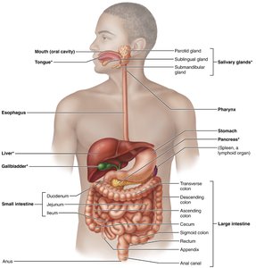
Major Digestive Processes
Six Essential Activities
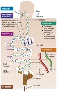
Food processing in the digestive system involves six key activities:
Ingestion: Taking in food.
Propulsion: Moving food through the canal, including swallowing (voluntary) and peristalsis (involuntary).
Mechanical breakdown: Physical breakdown of food (chewing, mixing, churning, segmentation).
Digestion: Chemical breakdown of food by enzymes into absorbable molecules.
Absorption: Passage of nutrients from the GI tract into blood or lymph.
Defecation: Elimination of indigestible residues as feces.
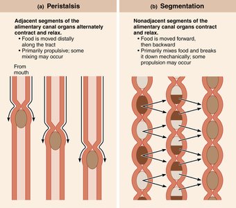
Peristalsis and Segmentation
These are two primary mechanisms for moving and mixing food in the GI tract:
Peristalsis: Alternating waves of contraction and relaxation propel food forward.
Segmentation: Local contractions mix food with digestive fluids, enhancing absorption.
Anatomy of the Digestive System
Peritoneum and Mesenteries
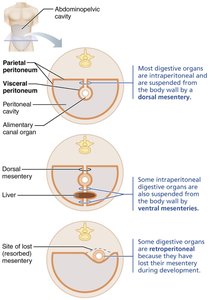
The peritoneum is a serous membrane lining the abdominopelvic cavity, consisting of:
Visceral peritoneum: Covers external surfaces of digestive organs.
Parietal peritoneum: Lines the body wall.
Peritoneal cavity: Space between membranes, filled with lubricating fluid.
Mesentery: Double layer of peritoneum that anchors organs and provides a route for vessels and nerves.
Intraperitoneal organs: Surrounded by peritoneum.
Retroperitoneal organs: Located behind the peritoneum.
Histology of the Alimentary Canal
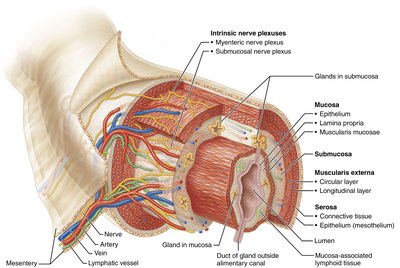
The alimentary canal wall has four basic layers (tunics):
Mucosa: Innermost layer; secretes mucus, enzymes, hormones; absorbs nutrients; protects against pathogens.
Submucosa: Areolar connective tissue with blood vessels, lymphatics, nerves, and elastic fibers.
Muscularis externa: Smooth muscle responsible for peristalsis and segmentation; includes circular and longitudinal layers.
Serosa: Outermost layer; visceral peritoneum. In retroperitoneal organs, replaced by adventitia.
Regulation of Digestive Activity
Enteric Nervous System
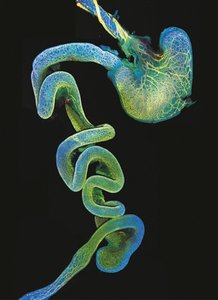
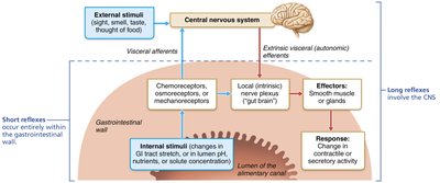
The GI tract has its own nervous system, the enteric nervous system (gut brain), which regulates digestive activities independently and in coordination with the central nervous system.
Intrinsic nerve plexuses: Submucosal and myenteric plexuses control motility.
Short reflexes: Mediated by enteric plexuses in response to local stimuli.
Long reflexes: Involve CNS and autonomic nerves, responding to internal and external stimuli.
Parasympathetic: Stimulates digestion; Sympathetic: Inhibits digestion.
The Mouth and Associated Organs
Anatomy of the Oral Cavity
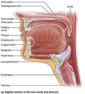
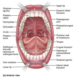
The mouth (oral cavity) is the entry point for food, bounded by lips, cheeks, palate, and tongue. It is lined with stratified squamous epithelium for protection.
Lips (labia): Orbicularis oris muscle covered with skin.
Cheeks: Buccinator muscles.
Palate: Hard (bony) and soft (muscular) palate; separates oral and nasal cavities.
Uvula: Fingerlike projection from soft palate.
Palatine tonsils: Located between palatoglossal and palatopharyngeal arches.
The Tongue
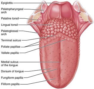
The tongue is a muscular organ that manipulates food, forms the bolus, initiates swallowing, and houses taste buds.
Intrinsic muscles: Change shape of tongue.
Extrinsic muscles: Alter tongue position.
Papillae: Filiform (friction, no taste buds), fungiform (taste), vallate (taste), foliate (taste).
Lingual tonsil: Immune function.
Salivary Glands
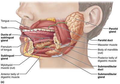
Salivary glands secrete saliva, which cleanses the mouth, moistens food, and begins starch digestion via amylase.
Major glands: Parotid, submandibular, sublingual.
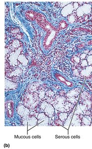
Serous cells: Produce watery, enzyme-rich secretion.
Mucous cells: Produce mucus.
Saliva composition: Water, electrolytes, amylase, lipase, mucin, lysozyme, IgA.
Teeth
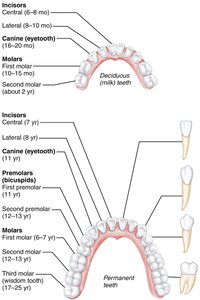
Teeth are housed in alveoli of the mandible and maxillae, and are essential for mastication (chewing).
Types: Incisors (cutting), canines (tearing), premolars and molars (grinding).
Dentition: Deciduous (20 baby teeth), permanent (32 adult teeth).
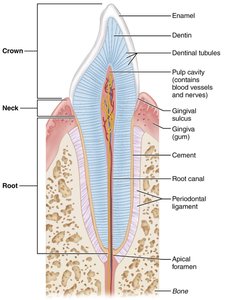
Structure: Crown (enamel), neck, root (cementum, periodontal ligament, dentin, pulp cavity).
The Pharynx and Esophagus
Pharynx
The pharynx connects the mouth to the esophagus and allows passage of food, fluids, and air. It is lined with stratified squamous epithelium and contains skeletal muscle layers for swallowing.
Esophagus
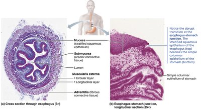
The esophagus is a muscular tube that transports food from the pharynx to the stomach. It is collapsed when not in use and features:
Mucosa: Stratified squamous epithelium.
Submucosa: Contains mucus-secreting glands.
Muscularis externa: Skeletal muscle superiorly, smooth muscle inferiorly.
Adventitia: Dense connective tissue.
Digestive Processes: Swallowing (Deglutition)
Swallowing Phases
Swallowing involves coordinated muscle actions and is divided into two phases:
Buccal phase: Voluntary, occurs in the mouth.
Pharyngeal-esophageal phase: Involuntary, controlled by the medulla and pons via the vagus nerve.
Summary Table: Layers of the Alimentary Canal
Layer | Main Components | Function |
|---|---|---|
Mucosa | Epithelium, lamina propria, muscularis mucosae | Secretion, absorption, protection |
Submucosa | Connective tissue, blood vessels, nerves | Support, elasticity, vascular supply |
Muscularis externa | Circular and longitudinal smooth muscle | Motility (peristalsis, segmentation) |
Serosa | Connective tissue, mesothelium | Protection, reduces friction |
Key Terms and Concepts
Peristalsis: Wave-like muscle contractions that move food along the GI tract.
Segmentation: Localized contractions that mix food with digestive juices.
Mesentery: Double layer of peritoneum anchoring organs and providing a pathway for vessels and nerves.
Enteric nervous system: Intrinsic neural network regulating GI motility and secretion.
Mucosa-associated lymphoid tissue (MALT): Immune defense in the GI tract.
Example: Clinical Relevance
Peritonitis: Inflammation of the peritoneum, often caused by infection or injury; can be life-threatening.
Heartburn: Pain from acid reflux into the esophagus; may indicate gastroesophageal reflux disease (GERD).
Additional info: These notes cover the foundational anatomy and physiology of the digestive system, including its structure, function, and regulatory mechanisms. For further study, review the processes of digestion and absorption, and the clinical conditions associated with digestive health.
