 뒤로
뒤로Chapter 23: The Respiratory System – Anatomy, Physiology, and Gas Exchange
스터디 가이드 - 스마트 노트
 자료에 맞춘 맞춤형 노트, 핵심 정의, 예시, 맥락을 확장해 제공합니다.
The Respiratory System
Overview and Organization
The respiratory system is responsible for the exchange of gases between the atmosphere and the body’s tissues. It is organized into upper and lower divisions, each with distinct anatomical structures and functions.
Upper Respiratory System: Includes the nose, nasal cavity, sinuses, and pharynx. These structures filter, warm, and moisten incoming air.
Lower Respiratory System: Comprises the larynx, trachea, bronchi, bronchioles, and lungs. These structures conduct air and facilitate gas exchange.
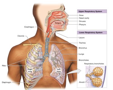
Key Terms and Definitions
Pharynx: Muscular tube connecting the nasal cavity to the larynx and esophagus; divided into nasopharynx, oropharynx, and laryngopharynx.
Larynx: Cartilaginous structure housing the vocal cords; functions in sound production and airway protection.
Bronchi: Airways branching from the trachea into the lungs.
Trachea: Windpipe; a tube reinforced by cartilage rings, conducting air to the bronchi.
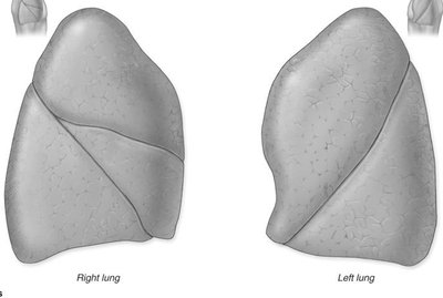
Lung: Organ of gas exchange, divided into lobes and covered by pleura.
Alveolus: Microscopic air sac where gas exchange occurs.
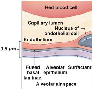
Respiratory membrane: Thin barrier between alveolar air and blood, facilitating gas diffusion.
Surfactant: Lipid-protein mixture reducing surface tension in alveoli, preventing collapse.
Anatomy of the Respiratory System
Upper Respiratory Tract
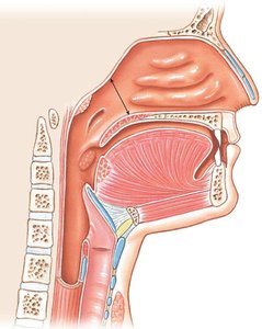
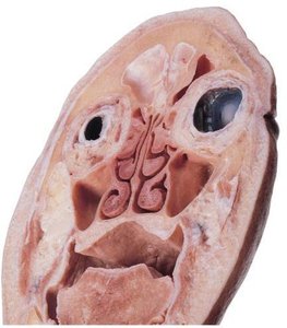
The upper respiratory tract prepares air for entry into the lungs by filtering, warming, and humidifying it. The nose and nasal cavity are lined by respiratory epithelium, which contains cilia and goblet cells to trap and move debris.
Nasal conchae: Increase surface area for air processing.
Hard and soft palate: Separate oral and nasal cavities.
The Pharynx
The pharynx is divided into three regions, each lined by different types of epithelium and serving distinct functions in respiration and digestion:
Nasopharynx: Posterior to nasal cavity; lined by pseudostratified ciliated columnar epithelium.
Oropharynx: Posterior to oral cavity; lined by stratified squamous epithelium.
Laryngopharynx: Posterior to larynx; lined by stratified squamous epithelium.
Lower Respiratory Tract
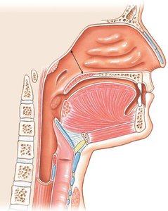
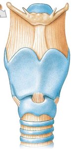
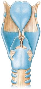
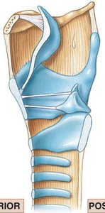
The lower respiratory tract includes the larynx, trachea, bronchi, and lungs. The larynx is composed of several cartilages, including the thyroid, cricoid, and epiglottis, which protect the airway and facilitate sound production.
Thyroid cartilage: Largest cartilage, forms the laryngeal prominence.
Cricoid cartilage: Forms the base of the larynx.
Epiglottis: Prevents food from entering the airway during swallowing.
The Trachea
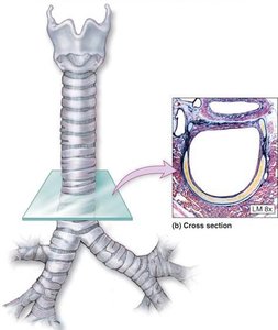
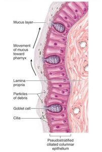
The trachea is a flexible tube supported by C-shaped cartilage rings. The trachealis muscle adjusts diameter for airflow regulation. The lining is pseudostratified ciliated columnar epithelium, which moves mucus upward toward the pharynx.
The Lungs and Pleura
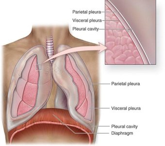
The lungs are located in the thoracic cavity, separated by the mediastinum, and covered by a double-layered serous membrane called the pleura. The pleural cavity contains fluid to reduce friction during breathing.
Parietal pleura: Lines the thoracic wall.
Visceral pleura: Covers the lung surface.
The Bronchial Tree
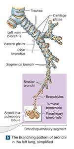
The bronchial tree consists of branching airways from the trachea to the alveoli. It is divided into the conducting zone (air movement) and the respiratory zone (gas exchange).
Primary bronchi: Enter each lung.
Secondary (lobar) bronchi: Supply lung lobes.
Tertiary (segmental) bronchi: Supply bronchopulmonary segments.
Bronchioles: Smallest airways leading to alveoli.
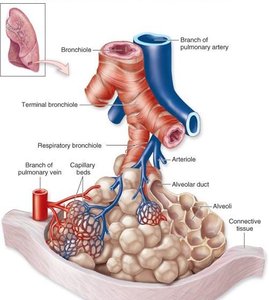
The Alveolus and Respiratory Membrane
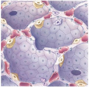
Alveoli are the sites of gas exchange. The respiratory membrane consists of three layers: squamous epithelial cells lining the alveolus, endothelial cells lining the capillary, and a fused basal lamina. Surfactant produced by Type II alveolar cells reduces surface tension, preventing alveolar collapse.
Type I alveolar cells: Form the structure of the alveolar wall.
Type II alveolar cells: Produce surfactant.
Respiratory Physiology
Processes of Respiration
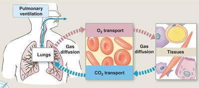
Respiration involves two integrated processes: external respiration (gas exchange between lungs and blood) and internal respiration (gas exchange between blood and tissues). The three steps are:
Pulmonary ventilation: Movement of air in and out of the lungs.
Gas diffusion: Exchange of O2 and CO2 across membranes.
Transport of O2 and CO2: Movement in blood to tissues.
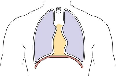
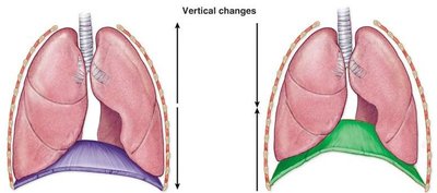
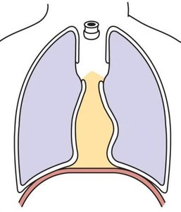
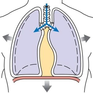
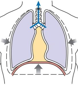
Pulmonary Ventilation: Mechanics of Breathing
Breathing is driven by pressure changes in the thoracic cavity. Boyle’s Law states that pressure and volume are inversely related: .
Inspiration: Diaphragm contracts, thoracic volume increases, pressure decreases, air enters lungs.
Expiration: Diaphragm relaxes, thoracic volume decreases, pressure increases, air exits lungs.
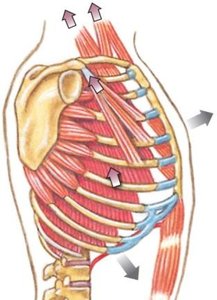
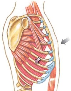
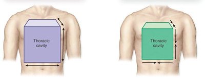

Gas Exchange and Partial Pressures
Gas exchange occurs across the respiratory membrane and is governed by partial pressures. Dalton’s Law states that the total pressure of a gas mixture is the sum of the partial pressures of individual gases: .
Partial pressure: The pressure exerted by a single gas in a mixture.
Gas diffusion: Gases move from areas of higher partial pressure to lower partial pressure.
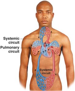
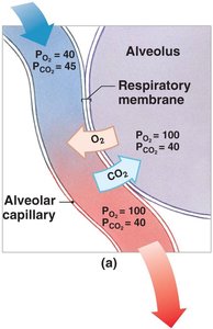
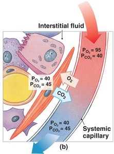
Transport of Oxygen and Carbon Dioxide
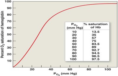
Oxygen is transported primarily bound to hemoglobin in red blood cells. The oxygen-hemoglobin saturation curve shows the relationship between partial pressure of oxygen (pO2) and hemoglobin saturation. Factors such as pH, temperature, and pCO2 affect this curve.
Oxygen: Bound to hemoglobin; saturation depends on pO2.
Carbon dioxide: Transported as dissolved gas, bound to hemoglobin, or as bicarbonate (via carbonic acid formation).
Factors Affecting Breathing
Breathing is regulated by plasma levels of CO2, O2, and pH. High plasma CO2 (hypercapnia) increases respiratory rate, while low plasma CO2 (hypocapnia) decreases it. Low blood oxygen (hypoxia) also stimulates breathing.
Hypercapnia: Elevated CO2 levels.
Hypocapnia: Reduced CO2 levels.
Hypoxia: Low O2 levels.
Anoxia: Absence of O2.
Summary Table: Major Structures and Functions
Structure | Function |
|---|---|
Nose/Nasal cavity | Filters, warms, moistens air |
Pharynx | Passage for air and food |
Larynx | Sound production, airway protection |
Trachea | Conducts air to bronchi |
Bronchi | Distribute air to lungs |
Alveoli | Site of gas exchange |
Pleura | Reduces friction, protects lungs |
Key Equations
Boyle’s Law:
Dalton’s Law:
Additional info:
These notes expand on brief lecture points to provide a comprehensive overview of respiratory anatomy and physiology, including definitions, examples, and key equations. The included images directly reinforce anatomical and physiological concepts for exam preparation.
