 뒤로
Chapter 8: The Nervous System – Essentials of Anatomy & Physiology Study Notes
스터디 가이드 - 스마트 노트
 자료에 맞춘 맞춤형 노트, 핵심 정의, 예시, 맥락을 확장해 제공합니다.
The Nervous System: Overview
Introduction to the Nervous System
The nervous system is the most complex organ system in the human body, responsible for rapid communication and coordination of bodily functions. It works alongside the endocrine system to maintain homeostasis, but its responses are faster and more brief.
Key Functions:
Monitors internal and external environments
Integrates sensory information
Coordinates voluntary and involuntary responses

Anatomical Divisions of the Nervous System
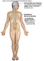
The nervous system is divided into two main anatomical divisions:
Central Nervous System (CNS): Includes the brain and spinal cord; integrates and coordinates sensory processing and motor transmission; site of higher functions (intelligence, memory, emotion).
Peripheral Nervous System (PNS): All nervous tissue outside the CNS; connects the CNS to the rest of the body.
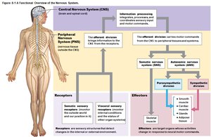
Functional Divisions of the PNS
Afferent Division: Brings information to the CNS from receptors in tissues and organs.
Efferent Division: Carries information from the CNS to effectors (muscles and glands).
Efferent Subdivisions:
Somatic Nervous System (SNS): Controls skeletal muscle contractions.
Autonomic Nervous System (ANS): Controls smooth and cardiac muscle, glands; includes sympathetic, parasympathetic, and enteric divisions.
Nervous Tissue
Cell Types in Nervous Tissue
Nervous tissue consists of two main cell types:
Neurons: Basic functional units; communicate with other cells.
Neuroglia (Glial Cells): Support and regulate the environment around neurons; more numerous and capable of division.
General Structure of Neurons
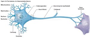
Neurons have specialized structures for communication:
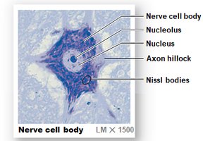
Cell Body: Contains nucleus, nucleolus, mitochondria, ribosomes, and rough ER (Nissl bodies).
Axon Hillock: Origin of action potentials.
Dendrites: Receive signals.
Axon: Transmits signals to other cells.
Neuron Regeneration
Most CNS neurons lack centrioles and cannot divide, making them irreplaceable if destroyed. Neural stem cells are mostly inactive except in specific regions (nose, hippocampus).
Structural Classification of Neurons
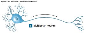
Multipolar Neurons: Two or more dendrites, one axon; most common in CNS; all motor neurons controlling skeletal muscles.
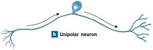
Unipolar Neurons: Dendrites and axon are continuous; cell body off to one side; most sensory neurons in PNS.
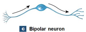
Bipolar Neurons: One dendrite, one axon; cell body between; rare, found in special sense organs.
Functional Classification of Neurons
Sensory (Afferent) Neurons: Receive information from sensory receptors and relay to CNS.
Motor (Efferent) Neurons: Carry instructions from CNS to effectors.
Interneurons: Located entirely within CNS; interconnect neurons; involved in higher functions (memory, learning).
Neuroglia (Glial Cells)
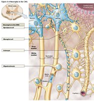
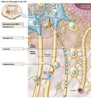
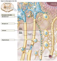
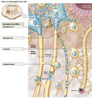
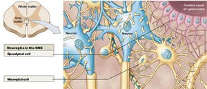
CNS Types: Astrocytes, oligodendrocytes, microglia, ependymal cells.
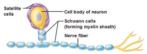
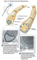
PNS Types: Satellite cells, Schwann cells.
Anatomical Organization of the Nervous System
Neuron Organization in the PNS and CNS
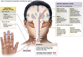
PNS: Collections of cell bodies (ganglia), bundles of axons (nerves), spinal and cranial nerves.
CNS: Collections of cell bodies (centers/nuclei), neural cortex, bundles of axons (tracts/columns), sensory (ascending) and motor (descending) pathways.
Membrane Potential and Action Potentials
The Membrane Potential
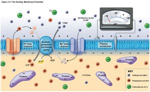
Living cells have a polarized membrane, with a resting membrane potential of –70 mV in neurons.
Factors: Imbalance in electrical charges, differing fluid composition (ECF high in Na+ and Cl–, ICF high in K+ and proteins).
Channels: Leak channels (always open), gated channels (open/close under specific conditions).
Maintaining Resting Membrane Potential
Sodium–Potassium Exchange Pump: Actively exchanges 3 Na+ out for 2 K+ in, maintaining the potential difference.
Changes in Membrane Potential
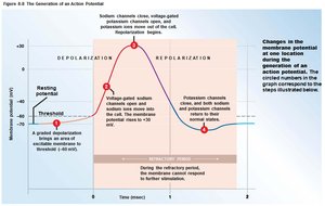
Depolarization: Opening Na+ channels shifts potential toward 0 mV.
Hyperpolarization: Opening K+ channels shifts potential more negative.
Graded Potentials
Local changes in membrane potential that decrease with distance; can trigger action potentials in excitable cells.
Action Potentials
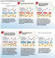
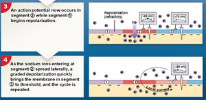
Propagated changes in membrane potential; follow the all-or-none principle; refractory period limits response rate.
Threshold: Level of depolarization required to initiate an action potential.
Propagation of Action Potentials
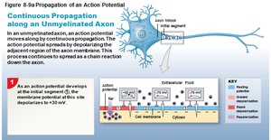
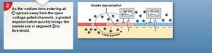
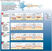
Continuous Propagation: Occurs in unmyelinated axons; relatively slow.
Saltatory Propagation: Occurs in myelinated axons; much faster.
Synapses and Neurotransmitters
The Synapse
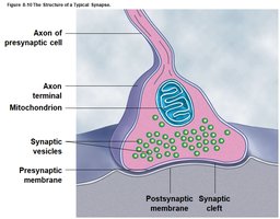
The synapse is the site of communication between neurons or between a neuron and another cell type.
Presynaptic Neuron: Sends signal; axon terminal contains neurotransmitter vesicles.
Postsynaptic Neuron: Receives signal; has receptors for neurotransmitters.
Acetylcholine and Other Neurotransmitters
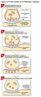
Acetylcholine (ACh): Released at cholinergic synapses; removed by acetylcholinesterase (AChE).
Other Neurotransmitters: Norepinephrine (NE), dopamine, GABA, serotonin, nitric oxide (NO), carbon monoxide (CO).
Excitatory vs. Inhibitory Neurotransmitters
Excitatory: ACh and NE trigger depolarization.
Inhibitory: Dopamine, GABA, serotonin trigger hyperpolarization.
Neuronal Pools
Groups of interconnected interneurons with specific functions; communicate in circuit patterns. 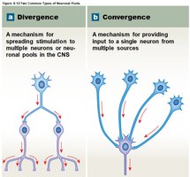
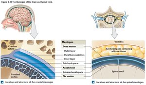
The Meninges
Three Layers of Meninges
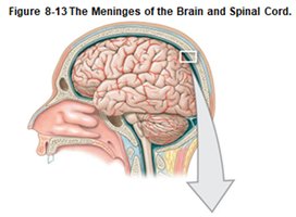
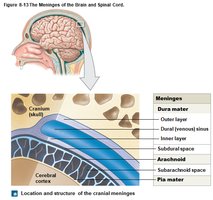
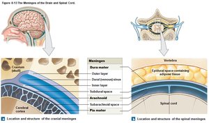
Specialized membranes surround the brain and spinal cord, providing stability and shock absorption.
Dura Mater: Tough, fibrous outer layer; contains dural folds and sinuses.
Arachnoid: Squamous epithelial cells; subarachnoid space filled with CSF.
Pia Mater: Innermost layer; highly vascularized, provides nutrients.
Spinal Cord Structure
Spinal Cord Overview
The spinal cord is the major neural pathway between the brain and PNS, integrating information and controlling reflexes.
Segments: 31 segments, each giving rise to a pair of spinal nerves.
Enlargements: Cervical and lumbosacral regions for limb innervation.
Gray and White Matter
Gray Matter: Contains neuron cell bodies; forms H or butterfly shape; projections called horns.
White Matter: Contains myelinated and unmyelinated axons; organized into columns (posterior, anterior, lateral).
Brain Structure and Function
Major Regions of the Brain
Cerebrum: Conscious thoughts, sensations, memory.
Diencephalon: Thalamus, hypothalamus, epithalamus; relay and processing centers.
Brainstem: Midbrain, pons, medulla oblongata; basic life functions.
Cerebellum: Motor coordination, balance.
Cerebral Cortex and Hemispheres
Gray Matter: Cerebral cortex (outer layer), basal nuclei (deeper).
White Matter: Deep to cortex, surrounds basal nuclei.
Gyri, Sulci, Fissures: Increase surface area.
Lobes: Frontal, parietal, temporal, occipital, insula.
Motor and Sensory Areas
Primary Motor Cortex: Precentral gyrus of frontal lobe.
Primary Sensory Cortex: Postcentral gyrus of parietal lobe.
Other Areas: Visual (occipital), auditory/olfactory (temporal), gustatory (frontal).
Association Areas and Processing Centers
Somatic Sensory Association: Recognizes touch.
Somatic Motor Association: Coordinates learned movements.
Wernicke’s Area: General interpretive area.
Broca’s Area: Speech center.
Basal Nuclei and Limbic System
Basal Nuclei: Subconscious control of muscle tone and learned movements.
Limbic System: Emotional states, memory storage/retrieval.
Diencephalon Components
Epithalamus: Contains pineal gland (melatonin).
Thalamus: Relay and processing center for sensory information.
Hypothalamus: Controls emotions, autonomic functions, hormone production.
Brainstem and Cerebellum
Midbrain: Visual and auditory processing, motor nuclei.
Pons: Links cerebellum with other brain regions; influences respiration.
Cerebellum: Adjusts motor activities, balance.
Medulla Oblongata: Connects brain to spinal cord; cardiovascular and respiratory centers.
Peripheral Nervous System (PNS)
Cranial and Spinal Nerves
Cranial Nerves: 12 pairs; originate from the brain; sensory, motor, or mixed functions.
Spinal Nerves: 31 pairs; connect to spinal cord; grouped by vertebral region.
Nerve Plexuses and Dermatomes
Plexuses: Cervical, brachial, lumbar, sacral; networks of major nerve trunks.
Dermatome: Region of skin monitored by a pair of spinal nerves; important for clinical diagnosis.
Reflexes and Pathways
Reflex Arc
Reflexes are rapid, automatic responses to stimuli, helping maintain homeostasis.
Monosynaptic Reflex: One synapse; e.g., patellar reflex.
Polysynaptic Reflex: Multiple synapses; e.g., withdrawal reflex.
Sensory and Motor Pathways
Sensory (Ascending) Pathways: Posterior column, spinothalamic, spinocerebellar; sensory homunculus maps.
Motor (Descending) Pathways: Corticospinal (pyramidal), medial/lateral (extrapyramidal); motor homunculus maps.
Autonomic Nervous System (ANS)
Structure and Function
Sympathetic Division: "Fight-or-flight"; increases alertness, heart rate, dilates pupils, decreases digestive activity.
Parasympathetic Division: "Rest-and-digest"; constricts pupils, increases digestive activity, decreases heart rate.
Dual Innervation: Both divisions affect the same organ, balancing responses.
Aging and Integration
Aging Effects on the Nervous System
Reduction in brain size and weight
Decrease in neuron number and synaptic connections
Decreased blood flow and neurotransmitter production
Increase in plaques; dementia may result
Integration with Other Body Systems
The nervous system interacts with all other body systems, coordinating responses and maintaining homeostasis.
Summary Table: Structural and Functional Divisions of the Nervous System
Division | Components | Main Functions |
|---|---|---|
Central Nervous System (CNS) | Brain, Spinal Cord | Integration, coordination, higher functions |
Peripheral Nervous System (PNS) | Nerves, Ganglia | Communication between CNS and body |
Somatic Nervous System (SNS) | Skeletal muscles | Voluntary control |
Autonomic Nervous System (ANS) | Cardiac/smooth muscle, glands | Involuntary regulation |
Sympathetic Division | Thoracic/lumbar spinal segments | Fight-or-flight responses |
Parasympathetic Division | Brainstem, sacral spinal segments | Rest-and-digest responses |
