 뒤로
Digestive System: Structure, Function, and Anatomy
스터디 가이드 - 스마트 노트
 자료에 맞춘 맞춤형 노트, 핵심 정의, 예시, 맥락을 확장해 제공합니다.
Digestive System Overview
Major Groups of Digestive Organs
The digestive system consists of two main groups: the alimentary canal (gastrointestinal tract) and accessory digestive organs. The alimentary canal is a continuous muscular tube extending from the mouth to the anus, responsible for digestion and absorption. Accessory organs assist in digestion through mechanical and chemical means.
Alimentary Canal: Includes the mouth, pharynx, esophagus, stomach, small intestine, and large intestine.
Accessory Digestive Organs: Teeth, tongue, gallbladder, salivary glands, liver, and pancreas.
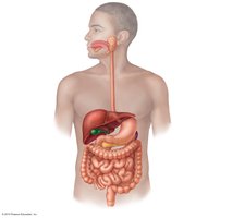
Digestive Processes
Six Essential Activities
The digestive system performs six essential activities to process food and absorb nutrients:
Ingestion: Intake of food into the mouth.
Mechanical Breakdown: Physical breakdown of food (chewing, churning, segmentation).
Propulsion: Movement of food through the tract (swallowing and peristalsis).
Digestion: Chemical breakdown of food into absorbable molecules.
Absorption: Passage of nutrients from the GI tract into the blood or lymph.
Defecation: Elimination of indigestible substances as feces.
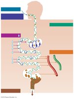
Peristalsis and Segmentation
Two primary movements facilitate food propulsion and mixing:
Peristalsis: Coordinated contraction and relaxation of adjacent segments of the alimentary canal, propelling food distally.
Segmentation: Alternating contraction of nonadjacent segments, mixing food and promoting absorption.
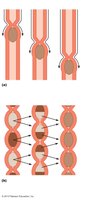
Peritoneum and Peritoneal Cavity
Structure and Function
The peritoneum is a serous membrane lining the abdominal cavity and covering abdominal organs. It consists of two layers:
Visceral Peritoneum: Covers external surfaces of most digestive organs.
Parietal Peritoneum: Lines the body wall.
Peritoneal Cavity: Space between the two peritoneums, containing lubricating fluid.
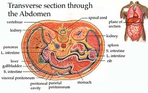
Mesentery and Omenta
The mesentery is a double layer of peritoneum that provides routes for blood vessels, lymphatics, and nerves, and helps anchor organs. The greater omentum is a visceral mesentery hanging from the stomach, involved in fat storage, immune function, and isolation of infection.
Retroperitoneal Organs: Located posterior to the peritoneum.
Intraperitoneal Organs: Surrounded by peritoneum.
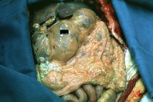
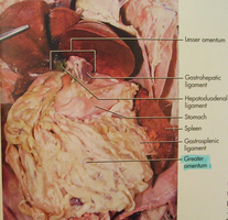
Homeostatic Imbalance: Peritonitis
Causes and Effects
Peritonitis is inflammation of the peritoneum, often caused by abdominal wounds, perforating ulcers, or ruptured appendix. Peritoneal coverings stick together to localize infection, but widespread peritonitis is dangerous and requires prompt treatment with antibiotics and debris removal.
Blood Supply: Splanchnic Circulation
Arterial and Portal Circulation
The digestive organs receive blood from branches of the aorta, including hepatic, splenic, and gastric arteries, as well as mesenteric arteries. The hepatic portal circulation drains nutrient-rich blood from digestive organs and delivers it to the liver for processing.
Histology of the Alimentary Canal
Four Basic Layers (Tunics)
The alimentary canal wall consists of four layers:
Mucosa: Innermost layer, involved in secretion and absorption.
Submucosa: Contains blood vessels, lymphatics, and nerves.
Muscularis Externa: Responsible for segmentation and peristalsis.
Serosa: Outermost layer, reduces friction.
Functional Anatomy: Mouth
Oral Cavity Structure
The oral cavity is bounded by lips, cheeks, palate, and tongue. It is lined with stratified squamous epithelium to withstand abrasion.
Lips and Cheeks: Contain muscles for facial expression and mastication.
Vestibule: Space between lips/cheeks and teeth/gums.
Labial Frenulum: Median attachment of each lip to gum.
Palate
Hard Palate: Bony, helps create friction against tongue.
Soft Palate: Muscular, closes off nasopharynx during swallowing; uvula projects from its edge.
Tongue
The tongue is a skeletal muscle organ involved in food manipulation, bolus formation, swallowing, speech, and taste. It is attached to the floor of the mouth by the lingual frenulum.
Intrinsic Muscles: Change shape of tongue.
Extrinsic Muscles: Alter tongue position.
Papillae: Filiform (friction, no taste buds), fungiform, circumvallate, foliate (contain taste buds).
Salivary Glands
Salivary glands produce saliva, which cleanses the mouth, dissolves food chemicals, moistens food, and begins starch digestion.
Extrinsic Glands: Parotid, submandibular, sublingual (produce most saliva).
Intrinsic Glands: Scattered throughout oral cavity.
Serous Cells: Produce watery, enzyme-rich fluid.
Mucous Cells: Produce mucus.
Teeth
Teeth are essential for mechanical digestion, tearing and grinding food. Humans have two sets: primary (deciduous) and permanent dentitions.
Incisors: Cutting.
Canines: Tearing.
Premolars: Grinding/crushing.
Molars: Best grinders.
Pharynx and Esophagus
Pharynx
The pharynx allows passage of food, fluids, and air. It is lined with stratified squamous epithelium and contains skeletal muscle layers for swallowing.
Esophagus
The esophagus is a muscular tube connecting the pharynx to the stomach. It passes through the diaphragm and joins the stomach at the cardial orifice, surrounded by the gastroesophageal sphincter.
Esophageal Mucosa: Stratified squamous epithelium, transitions to simple columnar at the stomach.
Muscularis Externa: Skeletal muscle superiorly, smooth muscle inferiorly.
Adventitia: Outermost connective tissue layer.
Digestive Processes: Mouth
Ingestion and Mastication
Food is ingested and mechanically digested by chewing. The tongue mixes food with saliva and forms a bolus. Mastication is partly voluntary and partly reflexive.
Deglutition (Swallowing)
Swallowing involves coordinated activity of the tongue, soft palate, pharynx, and esophagus, requiring 22 muscle groups. It consists of two phases:
Buccal Phase: Voluntary contraction of the tongue.
Pharyngeal-Esophageal Phase: Involuntary, controlled by the medulla and lower pons via the vagus nerve.
Stomach
Gross Anatomy
The stomach is a muscular organ that chemically digests food into chyme. It consists of the cardia, fundus, body, and pyloric part, which connects to the duodenum via the pyloric sphincter.
Microscopic Anatomy
Muscularis Externa: Three layers (longitudinal, circular, oblique) for churning and mixing.
Mucosa: Simple columnar epithelium, gastric pits, and glands producing gastric juice.
Gastric Glands
Mucous Neck Cells: Secrete acidic mucus.
Parietal Cells: Secrete HCl and intrinsic factor.
Chief Cells: Secrete pepsinogen and lipases.
Enteroendocrine Cells: Secrete hormones and paracrines.
Mucosal Barrier
The stomach is protected by a thick layer of bicarbonate-rich mucus, tight junctions between epithelial cells, and rapid cell turnover.
Small Intestine
Gross Anatomy
The small intestine is the major organ of digestion and absorption, extending from the pyloric sphincter to the ileocecal valve. It is subdivided into the duodenum, jejunum, and ileum.
Structural Modifications
Circular Folds (Plicae Circularis): Increase surface area.
Villi: Finger-like projections for absorption.
Microvilli: Brush border for increased absorption.
Intestinal Crypts
Secretory Cells: Produce intestinal juice.
Enteroendocrine Cells: Secrete hormones.
Paneth Cells: Secrete antimicrobial agents.
Stem Cells: Renew epithelium.
Liver and Gallbladder
Liver
The liver is the largest gland in the body, with four lobes. Its main digestive function is bile production, which emulsifies fats.
Gallbladder
The gallbladder stores and concentrates bile, releasing it into the duodenum as needed.
Pancreas
Structure and Function
The pancreas is both an endocrine and exocrine organ. It secretes insulin and glucagon (endocrine) and produces pancreatic juice containing digestive enzymes (exocrine).
Large Intestine
Gross Anatomy
The large intestine absorbs water and electrolytes, forming feces. It consists of the cecum, appendix, colon, rectum, and anal canal.
Teniae Coli: Bands of smooth muscle.
Haustra: Pocket-like sacs.
Epiploic Appendages: Fat-filled pouches.
Microscopic Anatomy
Mucosa: Simple columnar epithelium (except anal canal).
Goblet Cells: Abundant for mucus secretion.
Bacterial Flora
Bacteria in the colon synthesize vitamins, ferment indigestible carbohydrates, and release gases.
Summary Table: Digestive System Organs and Functions
Organ | Main Function | Key Features |
|---|---|---|
Mouth | Ingestion, mechanical digestion | Teeth, tongue, salivary glands |
Pharynx | Swallowing | Muscle layers, passage for food/air |
Esophagus | Propulsion | Muscular tube, sphincters |
Stomach | Chemical/mechanical digestion | Gastric glands, three muscle layers |
Small Intestine | Digestion, absorption | Villi, microvilli, circular folds |
Liver | Bile production | Four lobes, bile ducts |
Gallbladder | Bile storage | Muscular sac |
Pancreas | Enzyme secretion | Acini, ducts |
Large Intestine | Water absorption, feces formation | Haustra, teniae coli |
Key Equations and Concepts
Surface Area for Absorption:
pH of Gastric Juice:
Additional info:
Some images and anatomical details were inferred for completeness and clarity.
Tables and equations were expanded for academic context.
