 뒤로
Digestive System: Structure, Function, and Clinical Relevance
스터디 가이드 - 스마트 노트
 자료에 맞춘 맞춤형 노트, 핵심 정의, 예시, 맥락을 확장해 제공합니다.
Digestive System Overview
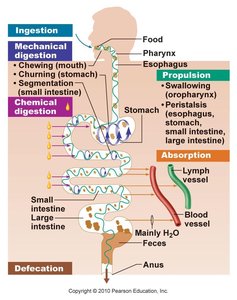
Digestive Functions
The digestive system is responsible for the breakdown and absorption of nutrients, as well as the elimination of waste. Its main functions include ingestion, mechanical and chemical digestion, absorption, and defecation.
Ingestion: Intake of food into the mouth.
Mechanical digestion: Physical breakdown of food (chewing, churning, segmentation).
Chemical digestion: Enzymatic breakdown of food molecules.
Absorption: Uptake of nutrients into blood or lymph.
Defecation: Elimination of undigested material.
Subdivisions of the Digestive System
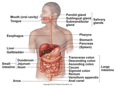
The digestive system consists of the digestive tract (GI tract) and accessory organs.
Digestive tract: A continuous tube from mouth to anus (~30 feet long).
Accessory organs: Teeth, tongue, liver, gallbladder, pancreas, and salivary glands aid in digestion.
Tissue Layers of the GI Tract
Histological Structure
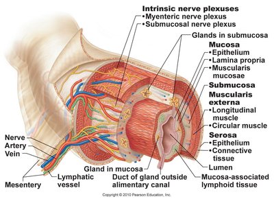
The GI tract wall is composed of four main layers, each with distinct functions.
Mucosa: Innermost layer; includes epithelium, lamina propria, and muscularis mucosae. Responsible for secretion and absorption.
Submucosa: Contains blood vessels, nerves, and glands.
Muscularis externa: Responsible for peristalsis and segmentation; typically has two layers (circular and longitudinal), but the stomach has three.
Serosa: Outermost layer; a serous membrane that reduces friction.
Peritoneum and Mesenteries
Serous Membranes
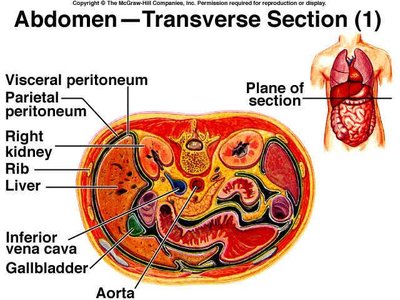
The peritoneum is a serous membrane lining the abdominal cavity and covering abdominal organs.
Visceral peritoneum: Covers organs.
Parietal peritoneum: Lines the abdominal wall.
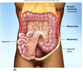
Mesentery: Suspends the small intestine and contains blood vessels, nerves, and lymphatics.
Mesocolon: Anchors the colon to the posterior body wall.
Omenta
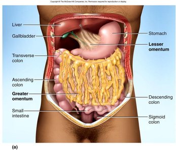
The omenta are folds of peritoneum that support and protect abdominal organs.
Lesser omentum: Attaches stomach to liver.
Greater omentum: Covers small intestines like an apron; contains adipose tissue, lymph nodes, blood vessels, and nerves.
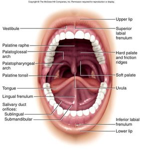
Mouth and Oral Cavity
Anatomical Features
The oral cavity is the entry point for food and is involved in mechanical digestion and speech.
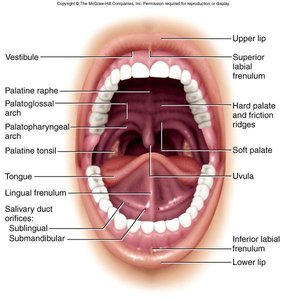
Cheeks and lips: Keep food between teeth; essential for chewing, speech, and suckling in infants.
Tongue: Muscular manipulator of food; contains papillae and taste buds.
Hard and soft palate: Separate oral and nasal cavities; aid in swallowing.
Uvula: Prevents food from entering the nasopharynx.
Dentition
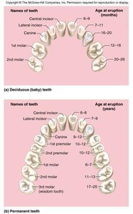
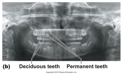
Teeth are essential for mechanical digestion. Humans have two sets of teeth:
Deciduous (baby) teeth: 20 teeth by age 2.
Permanent (adult) teeth: 32 teeth, erupt between ages 6 and 25.
Occlusal surfaces: The chewing surfaces; cusp numbers differ among teeth types.
Tooth Structure
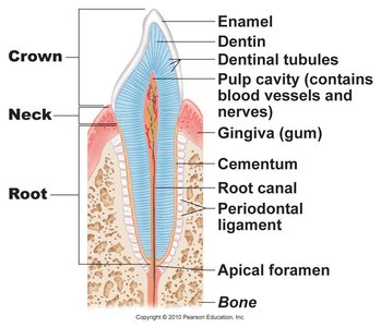
Each tooth consists of several distinct parts:
Enamel: Hard, noncellular secretion formed during development.
Cementum and dentin: Living tissues that support the tooth.
Pulp cavity: Contains nerves and blood vessels; extends into the root canal.
Gingiva (gum): Surrounds the base of the tooth.
Dental Hygiene
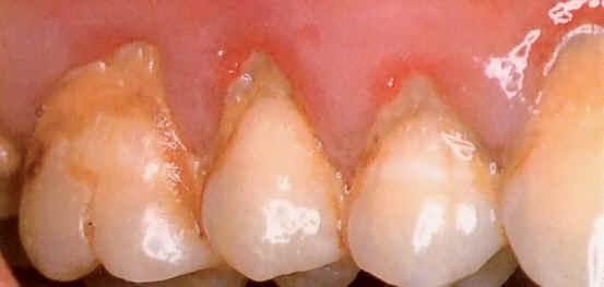
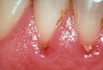
Proper dental hygiene is essential to prevent plaque, tartar, and gingivitis.
Plaque: Sticky film of bacteria and food debris.
Tartar: Hardened plaque.
Gingivitis: Inflammation of the gums.
Saliva and Salivary Glands
Functions of Saliva
Saliva is a hypotonic solution that aids in digestion and oral health.
Moistens food and begins starch and fat digestion.
Cleanses teeth and inhibits bacteria.
Binds food into a bolus for swallowing.
Contains enzymes: Salivary amylase (starch digestion), lingual lipase (fat digestion), lysozyme (kills bacteria), immunoglobulin A (inhibits bacterial growth).
Electrolytes: Na+, K+, Cl-, phosphate, bicarbonate.
pH: 6.8–7.0.
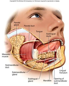
Salivary Glands
There are three pairs of major salivary glands:
Parotid gland
Submandibular gland
Sublingual gland
Salivation
1–1.5 L of saliva produced per day.
Salivation is stimulated by food, as well as sight, smell, and thought of food.
Salivatory nuclei in the brain coordinate secretion.
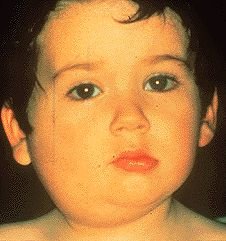
Mumps
Viral infection of the salivary glands.
Prevented by MMR vaccine.
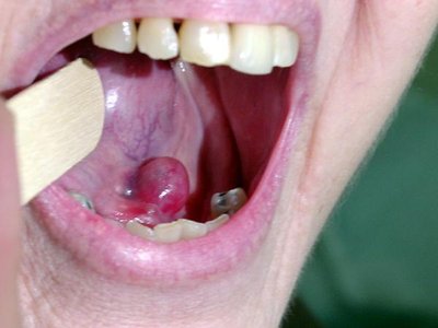
Salivary Gland Blockage
Blockage can cause swelling and pain.
Swallowing (Deglutition)
Process of Swallowing
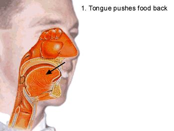
Swallowing is a complex process involving over 22 muscles, coordinated by the medulla and pons.
Voluntary phase: Tongue pushes food back into the pharynx.
Pharyngeal phase: Involuntary; food passes through pharynx.
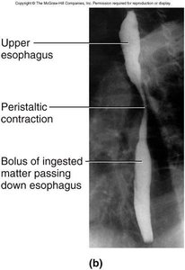
Esophageal phase: Peristaltic waves move food down the esophagus.
Liquids transit in ~2 seconds; food bolus may take ~8 seconds.
Stomach
Gross Anatomy and Function
The stomach is a J-shaped muscular sac primarily for food storage and initial digestion.
Internal volume: 50 mL–4 L.
Mechanical digestion: Churns and mixes food.
Chemical digestion: Begins protein and fat digestion.
Chyme: Soupy mixture resulting from digestion.
Absorption: Limited; absorbs aspirin and some lipid-soluble drugs.
Unique Features of Stomach Wall
Mucosa: Simple columnar glandular epithelium; contains tubular glands.
Muscularis externa: Three layers (longitudinal, circular, oblique).
Alkaline mucus: Protects stomach lining.
Cells of Gastric Glands
Mucous neck cells: Secrete mucus.
Parietal cells: Secrete HCl and intrinsic factor.
Chief cells: Secrete pepsinogen, chymosin, and lipase.
Gastric Secretions
HCl: Activates pepsin and lipase, breaks up tissues, liquefies food, antimicrobial.
Intrinsic factor: Essential for vitamin B12 absorption; necessary for RBC production.
Pepsin: Protein digestion; secreted as pepsinogen, activated by HCl.
Gastric Motility
Peristalsis occurs every 20 seconds.
Contractions mix food with gastric juice.
Stronger contractions at pyloric region eject small amounts of chyme.
Typical meal emptied in 4 hours; longer if fatty.
Vomiting
Induced by stomach stretching, psychological stimuli, or chemical irritants.
Emetic center in medulla coordinates retching and vomiting.
Frequent vomiting can cause electrolyte imbalances and tooth enamel decay.
Aspiration of stomach acid is dangerous for the respiratory tract.
Healthy Mucosa and Peptic Ulcer
Mucous protects stomach cells from acid.
Most peptic ulcers are caused by Helicobacter pylori.
Treatment is most successful with antibiotics.
Summary Table: Digestive System Functions
Function | Description | Location |
|---|---|---|
Ingestion | Intake of food | Mouth |
Mechanical Digestion | Physical breakdown | Mouth, stomach, small intestine |
Chemical Digestion | Enzymatic breakdown | Mouth, stomach, small intestine |
Absorption | Uptake of nutrients | Small intestine, large intestine |
Defecation | Elimination of waste | Rectum, anus |
Key Equations
pH calculation:
Additional info:
Digestive system disorders such as hiatal hernia, pyloric stenosis, and peptic ulcers are clinically significant and may require medical intervention.
Gastric bypass and lap band procedures are surgical treatments for obesity, altering stomach anatomy and function.
