 뒤로
Fluid, Electrolyte, and Acid-Base Balance: Structured Study Notes
스터디 가이드 - 스마트 노트
 자료에 맞춘 맞춤형 노트, 핵심 정의, 예시, 맥락을 확장해 제공합니다.
Fluid, Electrolyte, and Acid-Base Balance
Fluid Balance
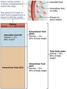
Fluid balance refers to the maintenance of the proper distribution and amount of water in the body. Water is essential for cellular function, and its distribution is regulated between two main compartments: intracellular and extracellular.
Intracellular Fluid (ICF): Fluid within cells, accounting for about 40% of body weight.
Extracellular Fluid (ECF): Fluid outside cells, including interstitial fluid and plasma, accounting for about 20% of body weight.
Adipose tissue contains little water, while lean tissue contains more.
Exchange of Gases, Nutrients, Water, and Wastes
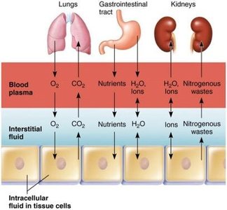
Substances such as oxygen, carbon dioxide, nutrients, water, ions, and wastes are exchanged between blood plasma, interstitial fluid, and intracellular fluid. This exchange is vital for homeostasis and cellular function.
Lungs: Exchange O2 and CO2 between blood and interstitial fluid.
Gastrointestinal tract: Absorbs nutrients and water into blood plasma.
Kidneys: Remove nitrogenous wastes and regulate ion balance.
Fluid Intake and Output
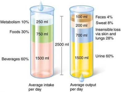
Fluid balance is achieved when daily water intake equals water output. Water intake comes from beverages, food, and metabolic processes, while output occurs via urine, feces, sweat, and insensible losses.
Average daily intake: 2500 mL (60% beverages, 30% food, 10% metabolism)
Average daily output: 2500 mL (60% urine, 28% insensible loss, 8% sweat, 4% feces)
Regulation of Water Intake
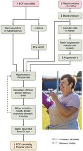
The hypothalamic thirst center controls the thirst mechanism, which is stimulated by osmoreceptors, dry mouth, decreased blood volume or pressure, and angiotensin II. Drinking water inhibits the thirst center through relief of dry mouth and activation of stretch receptors in the digestive tract.
Osmoreceptors: Detect changes in ECF osmolality.
Baroreceptors: Respond to changes in blood pressure.
Angiotensin II: Stimulates thirst in response to low blood volume.
Regulation of Water Output
Water output is regulated by obligatory losses (skin, airways, urine, sweat, feces) and is influenced by fluid intake, diet, and variable losses. The kidneys play a central role, beginning to eliminate excess water about 30 minutes after ingestion.
Minimum urine output: 500 mL/day to excrete wastes.
ADH (Antidiuretic Hormone): Regulates water reabsorption in the kidneys.
Influence of Antidiuretic Hormone (ADH)
ADH release increases water reabsorption in the collecting ducts, decreasing urine output and increasing body fluid volume. ADH is stimulated by increased ECF osmolality, decreased blood volume, and the renin-angiotensin-aldosterone system.
Low ADH: Fewer aquaporins, increased urine output.
High ADH: More aquaporins, decreased urine output.
Disorders of Water Balance
Dehydration
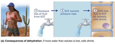
Dehydration results from excessive ECF fluid loss due to hemorrhage, burns, vomiting, diarrhea, sweating, water deprivation, diuretic abuse, or endocrine disturbances.
Early symptoms: Thirst, dry mouth, flushed skin, reduced urine output.
Severe effects: Weight loss, mental confusion, hypovolemic shock.
Hypotonic Hydration
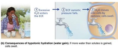
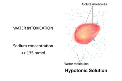
Hypotonic hydration occurs when excessive water intake dilutes ECF, causing water to move into cells by osmosis, leading to cell swelling and potentially cerebral edema.
Symptoms: Nausea, vomiting, muscular cramping, cerebral edema.
Treatment: Acute hyponatremia treated with hypertonic saline.
Edema
Edema is the accumulation of fluid in interstitial spaces, increasing the distance for diffusion of oxygen and nutrients and impairing tissue function. It is caused by factors that increase fluid flow out of blood.
Electrolyte Balance
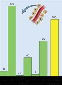
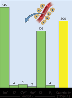
Electrolytes are salts that dissociate into ions in water and are essential for cellular function. Major cations include Na+, K+, Ca2+; major anions include Cl-, bicarbonate, and phosphate. Electrolyte balance is maintained by intestinal absorption and renal excretion.
Na+ and K+ Balance
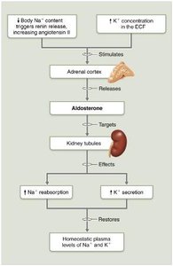
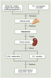
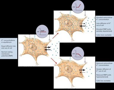
Sodium (Na+) is the most abundant cation in ECF and determines water distribution. Potassium (K+) is most abundant in ICF and is critical for maintaining resting membrane potential. Both are regulated by aldosterone, which promotes Na+ reabsorption and K+ excretion. Natriuretic peptides cause sodium and water excretion.


Regulation of Sodium Balance
Sodium balance is coupled to blood pressure and volume control. Aldosterone plays the largest role, reabsorbing most filtered Na+ in the proximal convoluted tubule (PCT) and nephron loops. When aldosterone is high, more Na+ is reabsorbed, increasing ECF volume; when low, more Na+ is excreted, decreasing ECF volume.
Na+ Imbalances
Hyponatremia: Sodium deficiency, often seen in athletes who lose sodium via sweating and replace fluids with plain water.
Hypernatremia: Excess sodium, less common.
Clinical Homeostatic Imbalance
Aldosterone deficiency (e.g., Addison's Disease) can result in large losses of NaCl and water, leading to hyponatremia and hypovolemia. Patients must be monitored and ingest adequate salt and fluids.
Regulation of Potassium Balance
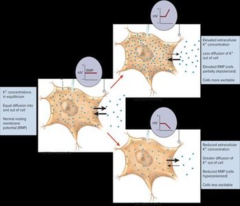
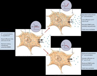
Potassium balance is regulated in the distal convoluted tubule (DCT) and collecting ducts by controlling the amount secreted into filtrate. The kidneys have limited ability to retain K+, so excessive loss can lead to hypokalemia.
K+ Imbalances
Hypokalemia: K+ deficiency, caused by chronic vomiting, diarrhea, laxative abuse, or aldosterone hypersecretion. Symptoms include muscle weakness and cardiac arrhythmia.
Hyperkalemia: Excess K+, caused by crush injury, aldosterone hyposecretion, or renal failure. Symptoms include muscle cramps, vomiting, diarrhea, and cardiac arrhythmia.
Acid-Base Balance
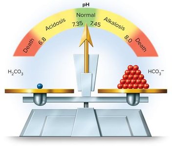
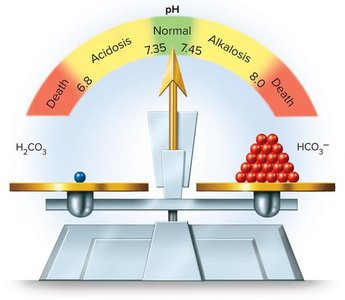
Acid-base balance is crucial for protein function and life processes. The normal blood pH range is 7.35–7.45. Alkalosis (pH > 7.45) causes hyperexcitability of nerve and muscle cells, while acidosis (pH < 7.35) depresses the nervous system and is more common.
Regulation of Acid-Base Balance
Chemical Buffers: Act quickly to stabilize pH by binding or releasing H+.
Respiratory System: Regulates CO2 levels, affecting pH.
Urinary System: Slowest, but most effective long-term regulator.
Chemical Buffers
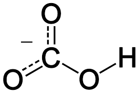
Buffers include bicarbonate, proteins, and phosphates. They can restore normal pH rapidly but are limited in capacity.
Respiratory Buffers
The respiratory system adjusts pH by changing the rate of CO2 exhalation. Excess CO2 causes acidosis (hypoventilation), while low CO2 causes alkalosis (hyperventilation).
Renal Buffers
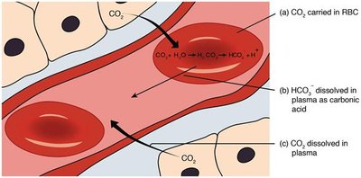
The kidneys regulate acid-base balance by generating bicarbonate and H+ from H2O and CO2. Bicarbonate is returned to the blood, raising pH, while H+ is excreted in urine.
Effects of Acidosis and Alkalosis
Blood pH below 6.8: Depresses CNS, can lead to coma and death.
Blood pH above 7.8: Overexcites nervous system, leading to muscle tetany, nervousness, convulsions, and death from respiratory arrest.
Acid-Base Imbalances in Relation to Electrolyte and Fluid Imbalances
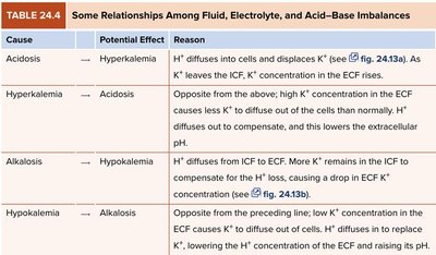
Acid-base imbalances often occur alongside fluid and electrolyte disturbances. For example, acidosis can cause hyperkalemia, while alkalosis can cause hypokalemia. The table below summarizes these relationships:
Cause | Potential Effect | Reason |
|---|---|---|
Acidosis | Hyperkalemia | H+ diffuses into cells and displaces K+; as K+ leaves the ICF, its concentration in the ECF rises. |
Hyperkalemia | Acidosis | High K+ concentration in the ECF causes less K+ to diffuse out of cells; H+ diffuses out to compensate, lowering ECF pH. |
Alkalosis | Hypokalemia | pH diffuses from ICF to ECF; more K+ remains in the ICF to compensate for H+ loss, causing a drop in ECF K+ concentration. |
Hypokalemia | Alkalosis | Low K+ concentration in the ECF causes K+ to diffuse out of cells; H+ diffuses in, lowering ECF K+ and raising its pH. |
Example: In diabetic ketoacidosis, acidosis leads to hyperkalemia, which can cause dangerous cardiac arrhythmias.
Additional info: These relationships are clinically important for understanding the effects of metabolic and respiratory disorders on electrolyte and fluid balance.
