 뒤로
Fundamentals of the Nervous System and Nervous Tissue
스터디 가이드 - 스마트 노트
 자료에 맞춘 맞춤형 노트, 핵심 정의, 예시, 맥락을 확장해 제공합니다.
Fundamentals of the Nervous System and Nervous Tissue
Overview of the Nervous System
The nervous system is a highly specialized network responsible for coordinating the body's activities by transmitting signals to and from different parts of the body. It is composed of two main cell types and is subdivided into several functional and anatomical regions.
Two Main Cell Types: Neurons (nerve cells) and neuroglia (supporting cells).
Subdivisions: Central Nervous System (CNS) and Peripheral Nervous System (PNS).
Subdivisions of the Nervous System
Central Nervous System (CNS): Consists of the brain and spinal cord; responsible for integrating, processing, and coordinating sensory data and motor commands.
Peripheral Nervous System (PNS): Includes all neural tissue outside the CNS; connects the CNS to limbs and organs.
Neurons and Neuroglia
Types of Cells
Neurons are the primary signaling cells, while neuroglia provide structural and metabolic support.
Neurons: Specialized for the conduction of electrical impulses.
Neuroglia: Support, protect, and nourish neurons.
Main Parts of a Neuron
Cell Body (Soma): Contains the nucleus and organelles.
Dendrites: Receive incoming signals.
Axon: Transmits electrical impulses away from the cell body.
Membrane Potentials and Ion Channels
Resting Membrane Potential
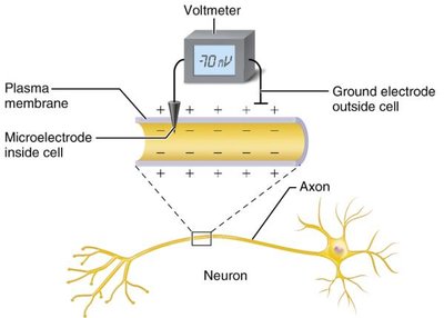
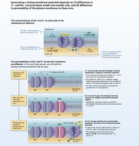
Neurons maintain a resting membrane potential, typically around -70 mV, due to differences in ion concentrations and membrane permeability.
Key Ions: Sodium (Na+) and Potassium (K+).
Mechanism: More K+ leaks out than Na+ leaks in, making the inside of the cell more negative.
Types of Ion Channels
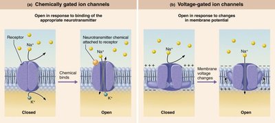
Leakage (Nongated) Channels: Always open, allowing ions to move along their gradients.
Gated Channels: Open or close in response to specific stimuli.
Chemically Gated (Ligand-Gated): Open with binding of a specific chemical (e.g., neurotransmitter).
Voltage-Gated: Open and close in response to changes in membrane potential.
Mechanically Gated: Open in response to physical deformation of the receptor.
Generation of Resting Membrane Potential
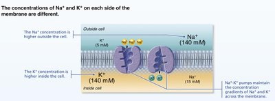
The resting membrane potential is established by the differential distribution of Na+ and K+ across the plasma membrane and the selective permeability of the membrane to these ions.
Na+: Higher concentration outside the cell.
K+: Higher concentration inside the cell.
Na+/K+ Pump: Maintains these gradients by pumping 3 Na+ out and 2 K+ in per ATP hydrolyzed.
Membrane Potential Changes and Signal Types
Graded Potentials
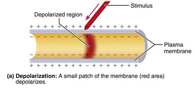
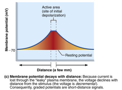
Graded potentials are short-distance, localized changes in membrane potential. They are essential for initiating action potentials.
Triggered by: Opening of gated ion channels.
Characteristics: Magnitude varies with stimulus strength; decays with distance.
Action Potentials
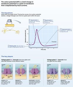
Action potentials are long-distance signals of axons, characterized by a rapid reversal of membrane potential.
All-or-None: Occur only if threshold is reached; do not decay with distance.
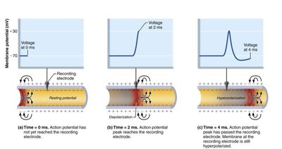
Phases: Resting state, depolarization, repolarization, hyperpolarization.
Phases of the Action Potential
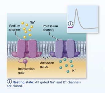
Resting State: All voltage-gated Na+ and K+ channels are closed.
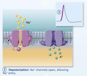
Depolarization: Na+ channels open, Na+ enters the cell, membrane potential becomes positive.
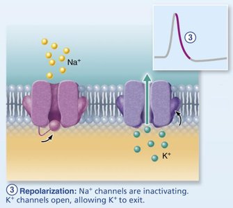
Repolarization: Na+ channels inactivate, K+ channels open, K+ exits the cell.
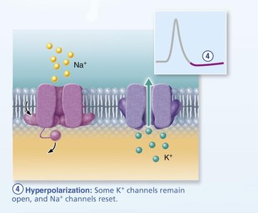
Hyperpolarization: Some K+ channels remain open, membrane potential dips below resting value.
Propagation and Refractory Periods
Propagation: Action potentials are self-propagating and travel in one direction along the axon.
Refractory Periods:
Absolute Refractory Period: No new action potential can be initiated.
Relative Refractory Period: Only a strong stimulus can initiate another action potential.
Synapses: Structure and Function
Types of Synapses
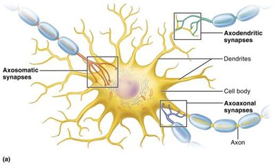
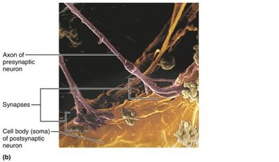
Synapses are specialized junctions where neurons communicate with other neurons or effector cells.
Electrical Synapses: Direct cytoplasmic connections via gap junctions; rapid, can be bidirectional.
Chemical Synapses: Use neurotransmitters to transmit signals across a synaptic cleft; unidirectional.
Neurotransmitters
Acetylcholine (ACh): Released at neuromuscular junctions and by some CNS and ANS neurons; degraded by acetylcholinesterase.
Catecholamines: Dopamine, norepinephrine, epinephrine; involved in stress response.
Indolamines: Serotonin (from tryptophan), histamine (from histidine); regulate mood, sleep, and other physiological processes.
Clinical Implications
Multiple Sclerosis (MS): Autoimmune destruction of CNS myelin, leading to impaired nerve conduction.
Local Anesthetics: Block voltage-gated Na+ channels, preventing action potentials.
Opioid Use: Synthetic and natural opiates can suppress breathing and cause dependency; naloxone can reverse overdose effects.
Summary Table: Ion Channel Types and Functions
Channel Type | Stimulus | Function | Example |
|---|---|---|---|
Chemically Gated | Binding of neurotransmitter | Initiates graded potentials | Synaptic transmission |
Voltage-Gated | Change in membrane potential | Generates and propagates action potentials | Axonal conduction |
Mechanically Gated | Physical deformation | Initiates receptor potentials in sensory neurons | Touch receptors |
Key Equations
Nernst Equation (for equilibrium potential of an ion):
Goldman-Hodgkin-Katz Equation (for membrane potential):
Additional info: These equations are fundamental for understanding how ion gradients and membrane permeability contribute to the resting membrane potential and action potentials in neurons.
