 뒤로
Fundamentals of the Nervous System and Nervous Tissue: Mini-Textbook Study Notes
스터디 가이드 - 스마트 노트
 자료에 맞춘 맞춤형 노트, 핵심 정의, 예시, 맥락을 확장해 제공합니다.
Fundamentals of the Nervous System and Nervous Tissue
Overview of the Nervous System
The nervous system is the master controlling and communicating system of the body. It is responsible for integrating sensory input, coordinating bodily functions, and facilitating rapid communication between different body regions.
Two main cell types: Neurons (excitable cells that transmit electrical signals) and neuroglia (supporting cells).
Subdivisions: Central Nervous System (CNS) and Peripheral Nervous System (PNS).
Main Parts of a Neuron
Neurons are highly specialized cells with distinct structural regions that facilitate their function in communication.
Cell body (soma): Contains the nucleus and metabolic machinery.
Dendrites: Receive incoming signals from other neurons.
Axon: Conducts electrical impulses away from the cell body toward other neurons or effectors.
Classification of Neurons
Neurons can be classified by structure and function, and are supported by major connective tissues.
Structural classification: Multipolar, bipolar, and unipolar neurons.
Functional classification: Sensory (afferent), motor (efferent), and interneurons.
Connective tissues: Endoneurium, perineurium, and epineurium surround nerve fibers, fascicles, and nerves, respectively.
Membrane Potentials and Ion Channels
Resting Membrane Potential
All cells have a resting membrane potential, but neurons can rapidly change this potential, making them highly excitable. The resting membrane potential of a typical neuron is about -70 mV, with the inside of the cell being more negative than the outside.
Generated by: Differences in ionic composition (mainly Na+ and K+) and differences in membrane permeability.
K+ plays the most important role in establishing the resting membrane potential.
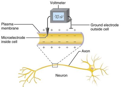
Role of Membrane Ion Channels
Ion channels are large proteins in the plasma membrane that allow specific ions to pass through, contributing to changes in membrane potential.
Leakage (nongated) channels: Always open, allowing ions to move down their concentration gradients.
Gated channels: Open or close in response to specific signals.
Chemically gated (ligand-gated) channels: Open with binding of a specific chemical (e.g., neurotransmitter).
Voltage-gated channels: Open and close in response to changes in membrane potential.
Mechanically gated channels: Open in response to physical deformation of the receptor.
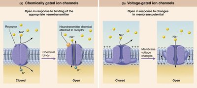
Electrochemical Gradients
The direction of ion movement is determined by the electrochemical gradient, which combines the concentration gradient and the electrical gradient.
Concentration gradient: Ions move from areas of higher to lower concentration.
Electrical gradient: Ions move toward areas of opposite electrical charge.
Establishing the Resting Membrane Potential
The resting membrane potential is established by the differential permeability of the membrane to Na+ and K+, and the activity of the Na+/K+ pump.
Membrane is more permeable to K+ than Na+.
K+ diffuses out of the cell more easily than Na+ diffuses in, making the inside more negative.
Na+/K+ ATPase pump maintains the gradients by pumping 3 Na+ out and 2 K+ in.
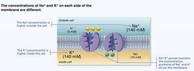
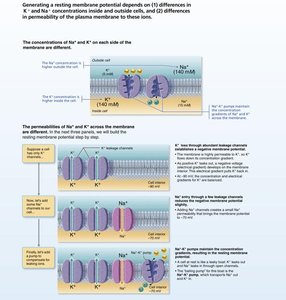
Graded Potentials and Action Potentials
Graded Potentials
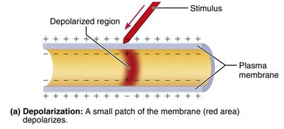
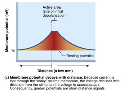
Graded potentials are short-distance, localized changes in membrane potential that occur in response to a stimulus. They are essential for initiating action potentials.
Triggered by the opening of gated ion channels.
Can result in depolarization or hyperpolarization.
Current flow dissipates quickly and decays with distance.
Action Potentials
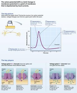
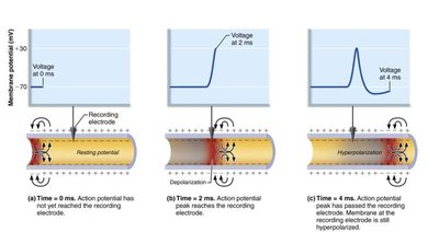
Action potentials (APs) are long-distance signals of axons, characterized by a brief reversal of membrane potential (from -70 mV to +30 mV). APs do not decay with distance and are the basis for nerve impulse transmission.
Occur only in excitable membranes (neurons and muscle cells).
Generated by the opening of specific voltage-gated channels.
All-or-none phenomenon: AP either happens completely or not at all.
Phases of the Action Potential
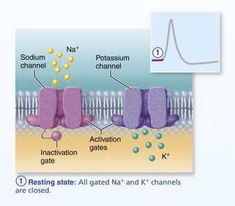
Resting State: All voltage-gated Na+ and K+ channels are closed. Only leakage channels are open, maintaining the resting membrane potential.
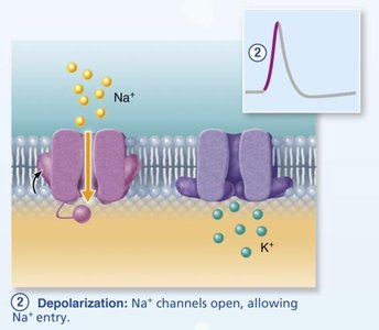
Depolarization: Voltage-gated Na+ channels open, Na+ rushes into the cell, and the membrane potential becomes less negative, reaching up to +30 mV.
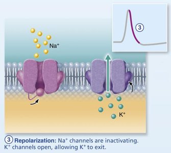
Repolarization: Na+ channels inactivate, and voltage-gated K+ channels open, allowing K+ to exit the cell, returning the membrane potential toward resting values.
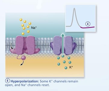
Hyperpolarization: Some K+ channels remain open, causing the membrane potential to dip below the resting value before returning to normal. Na+ channels reset.
Propagation and Refractory Periods
Once initiated, an action potential is self-propagating and moves in one direction along the axon. The refractory period ensures unidirectional propagation and limits the frequency of action potentials.
Absolute refractory period: No new AP can be generated.
Relative refractory period: Only a very strong stimulus can initiate another AP.
Factors Affecting Conduction Velocity
Axon diameter: Larger diameter fibers conduct faster.
Degree of myelination: Myelinated axons conduct impulses more rapidly via saltatory conduction.
Clinical Implications: Diseases like multiple sclerosis (MS) damage myelin sheaths, slowing or blocking nerve conduction. Local anesthetics block voltage-gated Na+ channels, preventing APs.
Synapses: Structure and Function
Types of Synapses
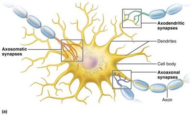
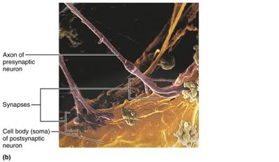
Synapses are specialized junctions where neurons communicate with other neurons or effector cells. There are two main types:
Electrical synapses: Direct cytoplasmic connections via gap junctions; allow rapid, bidirectional communication.
Chemical synapses: Use neurotransmitters to transmit signals across a synaptic cleft; ensure unidirectional communication.
Neurotransmitters
Neurotransmitters are the chemical messengers of the nervous system. They are classified by chemical structure and function.
Acetylcholine (ACh): Released at neuromuscular junctions and by some CNS and ANS neurons; degraded by acetylcholinesterase.
Catecholamines: Dopamine, norepinephrine, and epinephrine; involved in stress response.
Indolamines: Serotonin (from tryptophan) and histamine (from histidine); regulate mood, sleep, and other physiological processes.
Postsynaptic Potentials
Excitatory postsynaptic potentials (EPSPs): Depolarize the postsynaptic membrane, increasing the likelihood of an action potential.
Inhibitory postsynaptic potentials (IPSPs): Hyperpolarize the postsynaptic membrane, decreasing the likelihood of an action potential.
Clinical Implications: Opioids and Synaptic Transmission
Opioids, both natural and synthetic, act on the nervous system to alleviate pain but can lead to tolerance, dependence, and overdose. Naloxone is an opioid receptor antagonist used to reverse opioid overdose.
Summary Table: Ion Channel Types and Functions
Channel Type | Stimulus for Opening | Main Function |
|---|---|---|
Chemically gated | Binding of neurotransmitter | Initiate graded potentials |
Voltage-gated | Change in membrane potential | Generate and propagate action potentials |
Mechanically gated | Physical deformation | Detect sensory stimuli |
Key Equations
Nernst Equation (for equilibrium potential of an ion):
Goldman-Hodgkin-Katz Equation (for membrane potential):
Additional info: The above equations are fundamental for understanding how ion gradients and membrane permeability determine the resting and action potentials in neurons.
