뒤로
Fundamentals of the Nervous System and Nervous Tissue: Structured Study Notes
스터디 가이드 - 스마트 노트
 자료에 맞춘 맞춤형 노트, 핵심 정의, 예시, 맥락을 확장해 제공합니다.
Fundamentals of the Nervous System
Overview and Functions
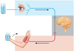
The nervous system is the master control and communication system of the body. It is responsible for monitoring internal and external changes, processing sensory input, and dictating motor responses. The three main functions are:
Sensory Input: Sensory receptors detect changes (stimuli) inside and outside the body, and this information is carried by afferent pathways.
Integration: The nervous system processes and interprets sensory input, determining the appropriate response.
Motor Output: The system activates effector organs (muscles or glands) via efferent pathways to produce a response.
Divisions of the Nervous System
Central and Peripheral Nervous Systems
The nervous system is divided into two main parts:
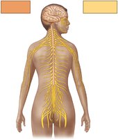
Central Nervous System (CNS): Composed of the brain and spinal cord. It serves as the integrating and command center, dictating motor output based on past experiences, reflexes, and current conditions.
Peripheral Nervous System (PNS): Consists of nerves extending from the brain and spinal cord (cranial and spinal nerves). Peripheral nerves link all regions of the body to the CNS. Ganglia are clusters of neuronal cell bodies in the PNS.
Sensory and Motor Subdivisions
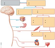
Sensory (afferent) signals are carried by nerve fibers of the PNS to the CNS, while motor (efferent) signals are carried away from the CNS to muscles and glands. These are further divided according to the region they serve:
Somatic body region
Visceral body region
This results in four main subdivisions:
Somatic sensory
Visceral sensory
Somatic motor
Visceral motor (autonomic nervous system)
Functional Subdivisions
Somatic and Visceral Sensory
Somatic sensory includes general senses (touch, pain, vibration, pressure, temperature) and proprioceptive senses (detect stretch in tendons and muscles, body position and movement). Special somatic senses include hearing, balance, and vision.
Visceral sensory includes general senses (stretch, pain, temperature, nausea, hunger) felt in internal organs, and special senses (taste, smell).
Somatic and Visceral Motor
Somatic motor signals contraction of skeletal muscles and is under voluntary control. Visceral motor regulates contraction of smooth and cardiac muscle and gland secretion, forming the autonomic nervous system (involuntary control).
Nervous Tissue
Cell Types
Nervous tissue is densely packed and intertwined, consisting of two main cell types:
Neurons: Transmit electrical signals.
Support cells (neuroglia): Nonexcitable, surround and wrap neurons.
Neuron Structure and Function
Neurons are the basic structural unit, specialized to conduct electrical impulses (action potentials). They exhibit longevity, do not divide (except neural stem cells), and have a high metabolic rate.
Cell body (soma): Contains organelles, chromatophilic substance (Nissl bodies), and neurofibrils for structural support.
Dendrites: Branching processes that transmit electrical signals toward the cell body; function as receptive sites.
Axon: Single impulse generator and conductor, transmits impulses away from the cell body; lacks chromatophilic bodies and protein synthesis.
Synapses
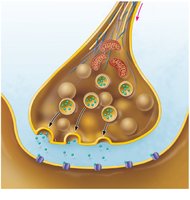
Synapses are sites where neurons communicate, mostly via chemical messengers (neurotransmitters) or electrically through gap junctions. The presynaptic neuron conducts signals toward the synapse, while the postsynaptic neuron transmits activity away. Types include axodendritic (most common) and axosomatic synapses.
Classification of Neurons
Structural Classification
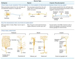
Neurons are classified by the number of processes extending from the cell body:
Multipolar: More than two processes; numerous dendrites and one axon.
Bipolar: Two processes; rare, found in special sensory organs.
Unipolar (pseudounipolar): One short, single process; common in sensory neurons.
Functional Classification
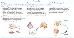
Neurons are also classified by the direction of impulse conduction relative to the CNS:
Sensory neurons: Transmit impulses toward the CNS; most are pseudounipolar.
Motor neurons: Carry impulses away from the CNS to effector organs; most are multipolar.
Interneurons: Lie between motor and sensory neurons; most are multipolar and confined to the CNS.
Neuroglia (Glial Cells)
Types and Functions
There are six types of neuroglia: four in the CNS and two in the PNS. They provide supportive functions, cover nonsynaptic regions, and insulate neurons.
Astrocytes (CNS): Most abundant; regulate neurotransmitter levels, blood flow, ionic environment, and neuronal growth.
Microglia (CNS): Defensive cells; phagocytes derived from monocytes.
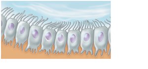
Ependymal cells (CNS): Line cerebrospinal fluid-filled cavities; bear cilia to circulate fluid.
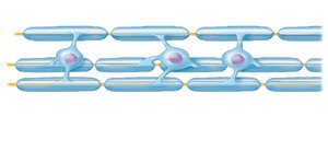
Oligodendrocytes (CNS): Form myelin sheaths around CNS axons.
Satellite cells (PNS): Surround neuron cell bodies in ganglia.
Schwann cells (PNS): Form myelin sheaths around PNS axons.
Myelin Sheaths
Structure and Function
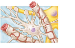
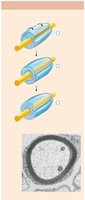
Myelin sheaths are segmented structures composed of the lipoprotein myelin, surrounding thicker axons. They form an insulating layer, prevent electrical leakage, increase impulse conduction speed, and make propagation more energy-efficient.
PNS: Formed by Schwann cells wrapping in concentric layers around the axon.
CNS: Formed by oligodendrocytes, which coil around several axons.
Gray and White Matter in the CNS
Gray Matter
Gray matter surrounds central cavities in the CNS, forming a butterfly-shaped region in the spinal cord. It contains neuron cell bodies, dendrites, short nonmyelinated neurons, and neuroglia. Synapses occur here.
White Matter
White matter lies external to gray matter, composed of myelinated axons and neuroglia. It contains no neuron cell bodies and consists of axons passing between CNS regions. Tracts are bundles of axons traveling to similar destinations.
Gross Anatomy of Nerves
Nerve Structure
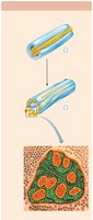
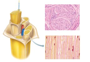
Nerves are cablelike organs in the PNS, consisting of numerous axons wrapped in connective tissue. Most nerves contain both myelinated and nonmyelinated sensory and motor axons.
Endoneurium: Surrounds Schwann cell-encased axon.
Perineurium: Surrounds nerve fascicle (bundle of axons).
Epineurium: Surrounds the whole nerve.
Neuronal Integration
CNS and PNS Interrelation
The CNS and PNS are functionally interrelated. Afferent PNS fibers respond to sensory stimuli, while efferent PNS fibers transmit motor stimuli from the CNS to muscles and glands. Interneurons in the CNS receive, direct, and transport information, and initiate motor responses.

Reflex Arcs
Components and Types
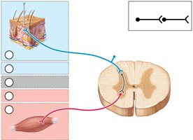
Reflex arcs are simple chains of neurons responsible for rapid, automatic motor responses. They consist of:
Receptor: Site where stimulus acts.
Sensory neuron: Transmits afferent impulses to CNS.
Integration center: One or more synapses in CNS.
Motor neuron: Conducts efferent impulses to effector.
Effector: Muscle or gland cell responding to impulses.
Monosynaptic and Polysynaptic Reflexes
Monosynaptic reflex: Simplest, fastest reflex with one synapse (e.g., knee-jerk reflex).
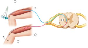
Polysynaptic reflex: More common, involves one or more interneurons (e.g., withdrawal reflex).
Neuronal Circuits and Processing
Types of Circuits
Diverging circuit: One presynaptic neuron synapses with several others.
Converging circuit: Many neurons synapse on a single postsynaptic neuron.
Reverberating circuit: Circuit receives feedback via collateral axon.
Serial processing: Signal passes along a single pathway to a specific destination.
Parallel processing: Input delivered along multiple pathways, resulting in multiple perceptions.
Neuronal Regeneration
Regeneration in PNS and CNS
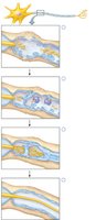
Neural injuries may cause permanent dysfunction. If only axons are destroyed, cell bodies may survive and axons can regenerate in the PNS, guided by Schwann cells. In the CNS, neuroglia do not form guiding bands and may hinder regeneration with inhibitory chemicals.
Nervous Tissue Development
Embryonic Development
The nervous system develops from the dorsal ectoderm, which invaginates to form the neural tube and neural crest. Neuroepithelial cells divide and become neuroblasts and neuroglial cells. Neural crest cells form sensory neurons, while neuroblasts form interneurons and motor neurons.