 뒤로
Fundamentals of the Nervous System and Nervous Tissue: Structured Study Notes
스터디 가이드 - 스마트 노트
 자료에 맞춘 맞춤형 노트, 핵심 정의, 예시, 맥락을 확장해 제공합니다.
Fundamentals of the Nervous System and Nervous Tissue
Characteristics and Functions of the Nervous System
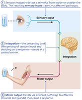
The nervous system is the master controlling and communicating system of the body. It uses electrical and chemical signals to rapidly and specifically coordinate bodily functions, often resulting in immediate responses.
Sensory Input: Information is gathered by sensory receptors about internal and external changes.
Integration: Processing and interpretation of sensory input, deciding on a response.
Motor Output: Activation of effectors (muscles and glands) produces a response.
Key Organs: Brain, spinal cord, nerves, and ganglia.
Organization of the Nervous System
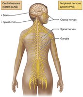
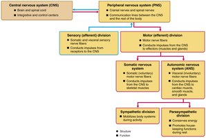
The nervous system is divided into two principal parts: the Central Nervous System (CNS) and the Peripheral Nervous System (PNS).
Central Nervous System (CNS): Consists of the brain and spinal cord. It is the integration and control center, interpreting sensory input and dictating motor output.
Peripheral Nervous System (PNS): Consists mainly of nerves that extend from the brain and spinal cord. It includes spinal nerves, cranial nerves, and the enteric nervous system (neurons in the walls of the gastrointestinal tract).
Functional Divisions of the PNS
Sensory (Afferent) Division: Somatic sensory fibers convey impulses from skin, skeletal muscles, and joints; visceral sensory fibers convey impulses from visceral organs.
Motor (Efferent) Division: Transmits impulses from CNS to effectors. Includes:
Somatic Nervous System: Voluntary control of skeletal muscles.
Autonomic Nervous System (ANS): Involuntary control of smooth muscle, cardiac muscle, and glands. Subdivided into:
Sympathetic Division: Mobilizes body systems during activity.
Parasympathetic Division: Conserves energy and promotes housekeeping functions during rest.
Histology of Nervous Tissue
Neuroglia (Glial Cells)
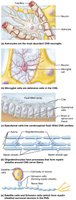
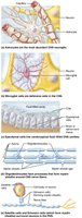
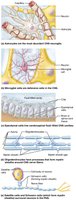
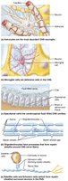
Nervous tissue consists of two principal cell types: neuroglia (glial cells) and neurons. Neuroglia are small cells that surround and wrap delicate neurons, providing support and protection.
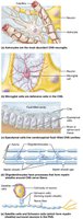
Astrocytes: Most abundant, versatile, and highly branched. Functions include supporting neurons, regulating exchanges between capillaries and neurons, guiding migration of young neurons, controlling the chemical environment, cleaning up leaked potassium ions, recycling neurotransmitters, and influencing neuronal functioning.
Microglial Cells: Small, ovoid cells with thorny processes. They monitor neurons, migrate toward injured neurons, and can transform to phagocytize microorganisms and neuronal debris.
Ependymal Cells: Range from squamous to columnar, may be ciliated. Line central cavities of the brain and spinal column, circulate cerebrospinal fluid (CSF), and form a permeable barrier between CSF and tissue fluid.
Oligodendrocytes: Branched cells with fewer processes. Their processes wrap CNS nerve fibers, forming insulating myelin sheaths.
Satellite Cells (PNS): Surround neuron cell bodies in the PNS, provide support, nourishment, and regulate the environment.
Schwann Cells (PNS): Surround all peripheral nerve fibers and form myelin sheaths around thicker nerve fibers. Vital to regeneration of damaged peripheral nerve fibers.
Neurons: Structure and Function
Neurons are the structural units of the nervous system. They are large, highly specialized cells that conduct nerve impulses. Neurons have extreme longevity, are mostly amitotic, and have a high metabolic rate requiring continuous oxygen and glucose.
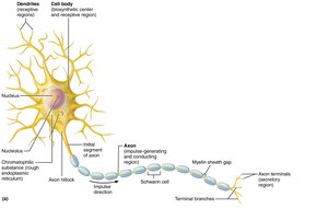
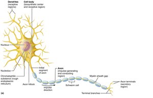
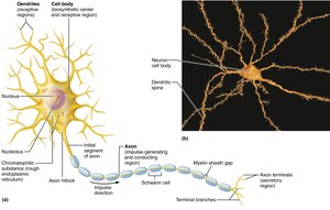
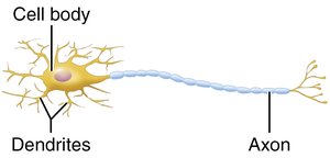
Cell Body (Soma): Contains the nucleus and nucleolus, surrounded by cytoplasm. It is the biosynthetic and metabolic center of the neuron.
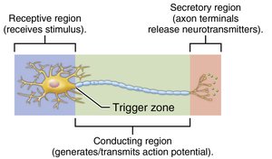
Dendrites: Receptive (input) regions that convey incoming messages toward the cell body as graded potentials.
Axon: Conducting region that generates and transmits nerve impulses away from the cell body. Axon terminals are the secretory region, releasing neurotransmitters.
Myelin Sheath
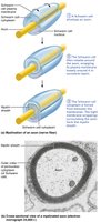
The myelin sheath is a white, fatty substance that coats many axons, especially long or large axons. It protects and electrically insulates axons, increasing the speed of nerve impulse transmission.
Myelination in the PNS: Formed by Schwann cells wrapping around the axon. Myelin sheath gaps (nodes of Ranvier) are sites where axon collaterals can emerge.
Myelination in the CNS: Formed by oligodendrocyte processes. Each cell can wrap up to 60 axons at once.
Classification of Neurons
Structural Classification
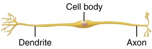
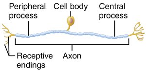
Neurons are classified by the number of processes extending from the cell body:
Multipolar Neurons: Many processes (1 axon, many dendrites). Major neuron type in CNS.
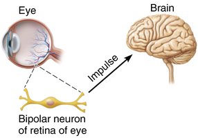
Bipolar Neurons: Two processes (1 axon, 1 dendrite). Rare, found in retina, ear, and olfactory mucosa.
Unipolar Neurons: One process (T-shaped, divides into two branches). Found mainly in PNS ganglia, functioning as sensory neurons.
Neuron Type | Structure | Location |
|---|---|---|
Multipolar | 1 axon, many dendrites | CNS (most abundant) |
Bipolar | 1 axon, 1 dendrite | Retina, ear, olfactory mucosa |
Unipolar | Single process, divides into two branches | PNS ganglia (sensory) |
Functional Classification
Neurons are also classified by the direction of impulse conduction:
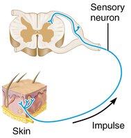
Sensory (Afferent) Neurons: Transmit impulses from sensory receptors toward CNS. Almost all are unipolar.
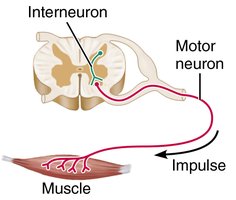
Motor (Efferent) Neurons: Carry impulses from CNS to effectors. Multipolar, most cell bodies in CNS.
Interneurons: Lie between motor and sensory neurons, shuttle signals through CNS pathways. Most abundant (99% of neurons).
Neuron Type | Function | Location |
|---|---|---|
Sensory | Impulse toward CNS | PNS ganglia |
Motor | Impulse from CNS to effectors | CNS |
Interneuron | Integration within CNS | CNS |
Membrane Potentials: Graded Potentials
Resting Membrane Potential
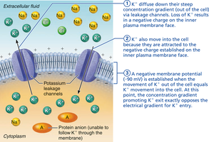
The plasma membrane of neurons has a resting membrane potential of approximately -70 mV, meaning the inside is slightly negative. This is primarily due to potassium ions (K+) moving out of the cell down their concentration gradient.
Graded Potentials
Graded potentials are short-lived, localized changes in membrane potential, usually occurring in dendrites or the soma. They are triggered by stimuli that open gated ion channels and can result in depolarization or hyperpolarization.
Amplitude: The stronger the stimulus, the greater the voltage change and the farther the current flows.
Decay: Graded potentials decay with time and distance.
Summation: Multiple graded potentials can add together (temporal or spatial summation) to reach threshold and trigger an action potential.
Types: Receptor potential (sensory neurons), postsynaptic potential (neurons), end-plate potential (muscle cells).
Membrane Potentials: Action Potentials
Action Potentials
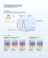
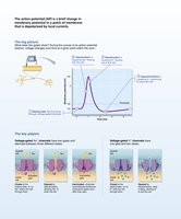
An action potential is a brief reversal of membrane potential, from -70 mV to +30 mV. It occurs only in excitable membranes (neurons and muscle cells) and does not decay over distance.
Phases of Action Potential
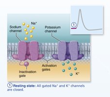
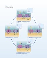
Resting State: All voltage-gated Na+ and K+ channels are closed. Only leakage channels are open.
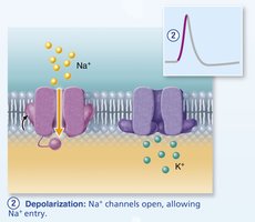
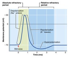
Depolarization: Voltage-gated Na+ channels open, Na+ rushes into the cell, causing the membrane to become less negative and reach +30 mV.
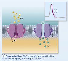
Repolarization: Na+ channels inactivate, K+ channels open, K+ exits the cell, returning the membrane potential toward resting.
Hyperpolarization: Some K+ channels remain open, causing the membrane potential to dip below resting.
Threshold and All-or-None Phenomenon
For an action potential to occur, depolarization must reach a threshold voltage. If threshold is reached, the action potential happens completely; if not, it does not occur at all.
Propagation of Action Potentials
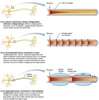
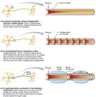
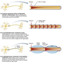
Action potentials are transmitted from their origin down the entire axon length toward terminals. In nonmyelinated axons, each segment depolarizes and repolarizes in sequence. In myelinated axons, propagation is faster (saltatory conduction).
Coding for Stimulus Intensity
All action potentials are alike and independent of stimulus intensity. The CNS distinguishes stimulus strength by the frequency of impulses.
Refractory Periods
Absolute Refractory Period: Time during which a neuron cannot trigger another action potential. Ensures one-way transmission.
Relative Refractory Period: Follows the absolute period; only exceptionally strong stimuli can trigger an action potential.
Conduction Velocity
Action potential conduction velocity depends on axon diameter and degree of myelination. Larger diameter and myelinated axons conduct impulses faster.
Continuous Conduction: Occurs in nonmyelinated axons, slower.
Saltatory Conduction: Occurs in myelinated axons, much faster.
Comparison: Graded Potentials vs. Action Potentials
Feature | Graded Potential | Action Potential |
|---|---|---|
Location | Dendrites, soma | Axon |
Amplitude | Variable, depends on stimulus | All-or-none, fixed |
Duration | Short-lived | Longer, does not decay |
Channel Types | Ligand-gated, mechanically-gated | Voltage-gated |
Function | Local signaling, integration | Long-distance signaling |
Additional info: These notes expand on the original lecture slides by providing definitions, examples, and structured tables for clarity. All images included directly reinforce the adjacent content and are strictly relevant to the explanation.
