 뒤로
Fundamentals of the Nervous System and Nervous Tissue – Study Notes
스터디 가이드 - 스마트 노트
 자료에 맞춘 맞춤형 노트, 핵심 정의, 예시, 맥락을 확장해 제공합니다.
Fundamentals of the Nervous System and Nervous Tissue
Overview of the Nervous System
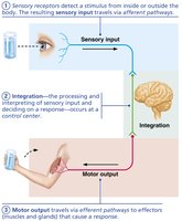
The nervous system is the master controlling and communicating system of the body. It uses electrical and chemical signals to coordinate rapid and specific responses to internal and external stimuli.
Sensory input: Information gathered by sensory receptors about changes inside and outside the body.
Integration: Processing and interpretation of sensory input, deciding what should be done at each moment.
Motor output: Activation of effector organs (muscles and glands) to produce a response.
Divisions of the Nervous System
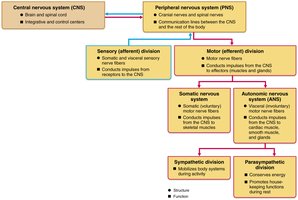
The nervous system is divided into two main parts:
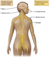
Central Nervous System (CNS): Consists of the brain and spinal cord. It is the integration and control center, interpreting sensory input and dictating motor output.
Peripheral Nervous System (PNS): Consists mainly of nerves extending from the brain and spinal cord (cranial and spinal nerves). It serves as communication lines between the CNS and the rest of the body. The PNS also includes the enteric nervous system in the gastrointestinal tract.
Functional Divisions of the PNS
Sensory (Afferent) Division: Transmits impulses from sensory receptors to the CNS. Includes somatic sensory fibers (from skin, muscles, joints) and visceral sensory fibers (from organs).
Motor (Efferent) Division: Transmits impulses from the CNS to effector organs. Subdivided into:
Somatic Nervous System: Voluntary control of skeletal muscles.
Autonomic Nervous System (ANS): Involuntary control of smooth muscle, cardiac muscle, and glands. The ANS is further divided into:
Sympathetic Division: Mobilizes body systems during activity ("fight or flight").
Parasympathetic Division: Conserves energy and promotes housekeeping functions during rest ("rest and digest").
Nervous Tissue: Neuroglia and Neurons
Neuroglia (Glial Cells)
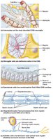
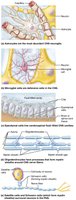
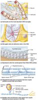
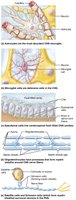
Neuroglia are supporting cells in nervous tissue. They provide structural and metabolic support to neurons, maintain the environment, and participate in signal transmission in the CNS and PNS.
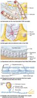
Astrocytes (CNS): Most abundant; support neurons, regulate the chemical environment, and help form the blood-brain barrier.
Microglial Cells (CNS): Act as phagocytes, removing debris and pathogens.
Ependymal Cells (CNS): Line brain ventricles and spinal cord central canal; circulate cerebrospinal fluid (CSF).
Oligodendrocytes (CNS): Form myelin sheaths around CNS axons.
Satellite Cells (PNS): Surround neuron cell bodies in ganglia; similar function to astrocytes.
Schwann Cells (PNS): Form myelin sheaths around PNS axons; vital for nerve fiber regeneration.
Neurons (Nerve Cells)
Neurons are the structural and functional units of the nervous system. They are specialized for the conduction of electrical impulses.
Longevity: Can live a lifetime.
Amitotic: Most do not divide after development.
High Metabolic Rate: Require continuous oxygen and glucose supply.
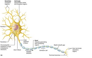
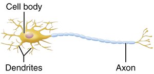
Structure: Consist of a cell body (soma) and processes (dendrites and axon).
Neuron Cell Body (Soma)
Contains the nucleus and organelles.
Acts as the biosynthetic and metabolic center.
Clusters in CNS are called nuclei; in PNS, ganglia.
Neuron Processes
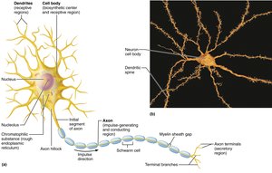
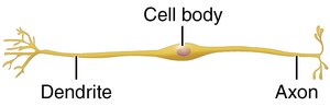
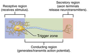
Dendrites: Short, branched processes that receive signals and convey them toward the cell body as graded potentials.
Axon: Single long process that transmits impulses away from the cell body. Axons may branch (axon collaterals) and end in axon terminals, which release neurotransmitters.
Bundles of axons in CNS are called tracts; in PNS, nerves.
Myelin Sheath
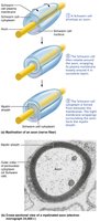
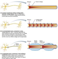
The myelin sheath is a white, fatty covering that insulates axons and increases the speed of impulse transmission.
PNS: Formed by Schwann cells; gaps between cells are called nodes of Ranvier.
CNS: Formed by oligodendrocyte processes; one cell can myelinate multiple axons.
Nonmyelinated fibers conduct impulses more slowly.
Classification of Neurons
Structural Classification
Multipolar: One axon, many dendrites (most common in CNS).
Bipolar: One axon, one dendrite (rare; found in retina, olfactory mucosa).
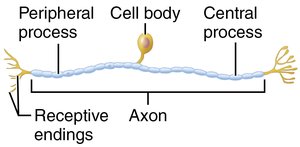
Unipolar (Pseudounipolar): Single short process that splits into peripheral and central processes (sensory neurons in PNS).
Type | Structure | Location |
|---|---|---|
Multipolar | Many dendrites, one axon | CNS, motor neurons |
Bipolar | One dendrite, one axon | Retina, olfactory epithelium |
Unipolar | Single process splits into two | Sensory neurons in PNS |
Functional Classification
Sensory (Afferent) Neurons: Transmit impulses from sensory receptors toward the CNS; mostly unipolar.
Motor (Efferent) Neurons: Carry impulses from the CNS to effectors; multipolar.
Interneurons (Association Neurons): Lie between sensory and motor neurons; most abundant; found entirely within the CNS.
Membrane Potentials and Electrical Signals
Basic Principles of Electricity
Voltage (V): Measure of potential energy generated by separated charge; measured in volts (V) or millivolts (mV).
Current (I): Flow of electrical charge between two points.
Resistance (R): Hindrance to charge flow.
Ohm's Law:
Ion Channels in the Plasma Membrane
Leakage (Nongated) Channels: Always open.
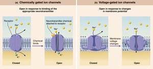
Gated Channels: Open or close in response to specific signals:
Chemically gated (ligand-gated): Open with binding of a specific chemical (e.g., neurotransmitter).
Voltage-gated: Open in response to changes in membrane potential.
Mechanically gated: Open in response to physical deformation.
Resting Membrane Potential
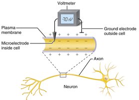
All cells have a resting membrane potential, but neurons can rapidly change it. The resting membrane potential is typically around -70 mV (inside negative relative to outside).
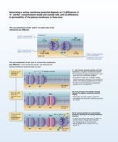
Generated by differences in ionic composition of intracellular fluid (ICF) and extracellular fluid (ECF), and differences in membrane permeability.
Potassium (K+) plays the most important role in generating the resting potential.
Changes in Membrane Potential
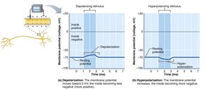
Depolarization: Decrease in membrane potential (inside becomes less negative); increases probability of producing a nerve impulse.
Hyperpolarization: Increase in membrane potential (inside becomes more negative); decreases probability of producing a nerve impulse.
Action Potentials
Generation and Propagation of Action Potentials
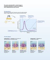
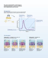
An action potential (AP) is a brief reversal of membrane potential that travels along the axon. It is the principal means of long-distance neural communication.
Occurs only in excitable membranes (neurons and muscle cells).
Does not decay with distance.
Involves opening of voltage-gated ion channels.
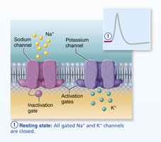
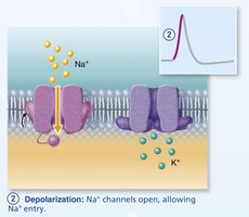
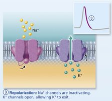
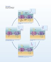
Phases of the Action Potential
Resting State: All voltage-gated Na+ and K+ channels are closed.
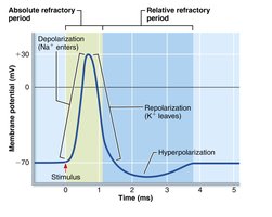
Depolarization: Na+ channels open, Na+ enters the cell, membrane potential becomes positive.
Repolarization: Na+ channels inactivate, K+ channels open, K+ exits the cell, membrane potential returns to negative.
Hyperpolarization: Some K+ channels remain open, causing a slight dip below resting potential.
Refractory Periods
Absolute Refractory Period: Neuron cannot trigger another AP; ensures one-way transmission.
Relative Refractory Period: Follows absolute; only a strong stimulus can trigger another AP.
Conduction Velocity
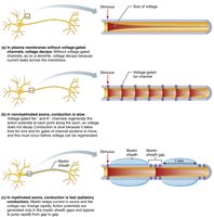
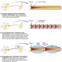
Depends on axon diameter (larger = faster) and degree of myelination.
Continuous conduction: Occurs in nonmyelinated axons; slower.
Saltatory conduction: Occurs in myelinated axons; AP "jumps" from node to node; much faster.
Synapses
Structure and Function of Synapses
Synapses are junctions that mediate information transfer from one neuron to another or to an effector cell. They ensure unidirectional communication and can be electrical or chemical.
Presynaptic neuron: Conducts impulses toward the synapse.
Postsynaptic neuron: Transmits impulses away from the synapse.
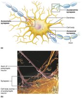
Types of synaptic connections: axodendritic, axosomatic, axoaxonal, dendrodendritic, somatodendritic.
Electrical Synapses
Neurons are electrically coupled via gap junctions.
Allow rapid, bidirectional communication; less common than chemical synapses.
Chemical Synapses
Most common type; specialized for release and reception of neurotransmitters.
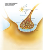
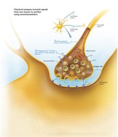
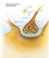
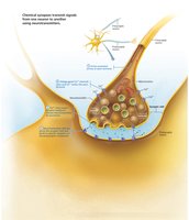
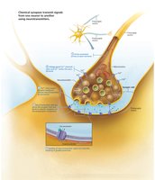
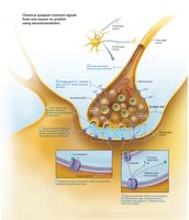
Composed of presynaptic axon terminal (with synaptic vesicles) and postsynaptic receptor region, separated by a synaptic cleft.
Transmission involves conversion of electrical signal to chemical (neurotransmitter release), then back to electrical in the postsynaptic cell.
Steps in Chemical Synaptic Transmission
AP arrives at axon terminal of presynaptic neuron.
Voltage-gated Ca2+ channels open; Ca2+ enters axon terminal.
Ca2+ entry causes synaptic vesicles to release neurotransmitter by exocytosis.
Neurotransmitter diffuses across synaptic cleft and binds to receptors on postsynaptic membrane.
Binding opens ion channels, creating graded potentials (excitatory or inhibitory).
Neurotransmitter effects are terminated by reuptake, enzymatic degradation, or diffusion away from the synapse.
Example: Acetylcholine is released at neuromuscular junctions, binds to receptors on muscle cells, and is quickly degraded by acetylcholinesterase.
Additional info: This guide covers the core concepts of nervous system structure and function, neuroglia, neuron anatomy, membrane potentials, action potentials, and synaptic transmission, as outlined in a typical Anatomy & Physiology curriculum.
