뒤로
Fundamentals of the Nervous System and Nervous Tissue – Study Notes
스터디 가이드 - 스마트 노트
 자료에 맞춘 맞춤형 노트, 핵심 정의, 예시, 맥락을 확장해 제공합니다.
Fundamentals of the Nervous System and Nervous Tissue
Overview of the Nervous System
The nervous system is the master control and communication system of the body. It is responsible for monitoring changes, processing information, and dictating responses. These functions are essential for maintaining homeostasis and coordinating bodily activities.
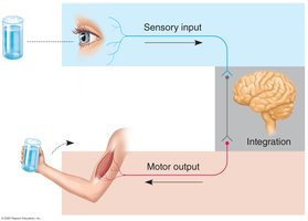
Sensory Input: Sensory receptors detect changes (stimuli) inside and outside the body and relay this information to the nervous system.
Integration: The nervous system processes and interprets sensory input, determining the appropriate response.
Motor Output: The nervous system activates effector organs (muscles and glands) to carry out a response.
Divisions of the Nervous System
The nervous system is divided into two main anatomical and functional subdivisions:
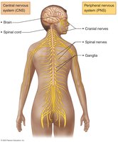
Central Nervous System (CNS): Consists of the brain and spinal cord. It is the integrating and command center of the nervous system.
Peripheral Nervous System (PNS): Composed of cranial nerves, spinal nerves, and ganglia. It links all regions of the body to the CNS.
Sensory Input and Motor Output Organization
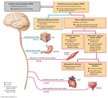
Sensory (afferent) signals are carried by PNS nerve fibers to the CNS, while motor (efferent) signals are carried away from the CNS to muscles and glands. These pathways are further divided based on the body region they serve:
Somatic Sensory: General senses (touch, pain, vibration, pressure, temperature) and special senses (hearing, balance, vision).
Visceral Sensory: Senses from internal organs (stretch, pain, temperature, nausea, hunger) and special senses (taste, smell).
Somatic Motor: Voluntary control of skeletal muscles.
Visceral Motor (Autonomic Nervous System): Involuntary control of smooth muscle, cardiac muscle, and glands.
Nervous Tissue
Cell Types in Nervous Tissue
Nervous tissue is composed of two main cell types:
Neurons: Excitable cells that transmit electrical signals (action potentials).
Neuroglia (Glial Cells): Nonexcitable support cells that surround and protect neurons.
Structure of a Typical Neuron
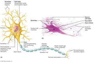
Neurons are the basic structural and functional units of the nervous system. They have specialized structures for receiving, integrating, and transmitting information.
Cell Body (Soma): Contains the nucleus and organelles; site of metabolic activity.
Dendrites: Branching processes that receive signals and transmit them toward the cell body.
Axon: Single, long process that transmits impulses away from the cell body to other neurons or effectors.
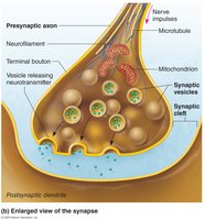
Axon Terminals (Terminal Boutons): Release neurotransmitters to communicate with other cells.
Synapses
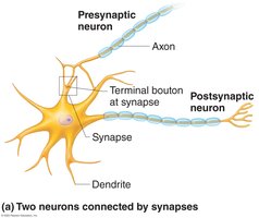
Synapses are specialized junctions where neurons communicate with other neurons, muscle cells, or glands. Most synapses use chemical messengers (neurotransmitters), but some are electrical (gap junctions).
Presynaptic Neuron: Conducts signal toward the synapse.
Postsynaptic Neuron: Receives the signal.
Synaptic Cleft: Small gap separating the two neurons.
Classification of Neurons
Neurons are classified structurally and functionally:
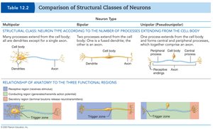
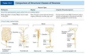
Multipolar: Many dendrites, one axon (most common type in CNS).
Bipolar: One dendrite, one axon (rare; found in special sensory organs).
Unipolar (Pseudounipolar): Single process that splits into two branches (sensory neurons in PNS).
Neuron Type | Structure | Location |
|---|---|---|
Multipolar | Many dendrites, one axon | Major neuron type in CNS |
Bipolar | One dendrite, one axon | Special sensory organs (retina, olfactory epithelium) |
Unipolar | Single process splits into two | Sensory neurons in PNS |
Functional Classification of Neurons
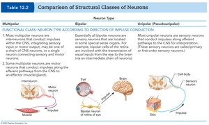
Sensory (Afferent) Neurons: Transmit impulses toward the CNS; mostly unipolar.
Motor (Efferent) Neurons: Carry impulses away from the CNS to effectors; mostly multipolar.
Interneurons: Lie between sensory and motor neurons; confined to the CNS; mostly multipolar.
Neuroglia (Glial Cells)
Neuroglia in the CNS
There are four types of neuroglia in the CNS:
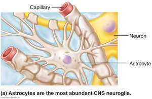
Astrocytes: Most abundant; regulate neurotransmitter levels, blood flow, and ionic environment; support neuronal growth and memory.
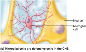
Microglia: Phagocytic cells; act as macrophages to remove debris and pathogens.
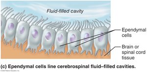
Ependymal Cells: Line central cavities of brain and spinal cord; circulate cerebrospinal fluid with cilia.
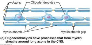
Oligodendrocytes: Form myelin sheaths around CNS axons.
Neuroglia in the PNS
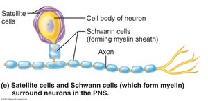
Satellite Cells: Surround neuron cell bodies in ganglia; regulate environment around neurons.
Schwann Cells (Neurolemmocytes): Surround axons in the PNS; form myelin sheaths.
Myelin Sheaths
Myelin Sheaths in the PNS
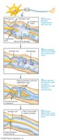
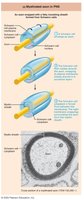
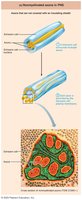
Myelin sheaths are segmented, insulating layers around axons that increase the speed of nerve impulse conduction. In the PNS, they are formed by Schwann cells wrapping around axons.
Myelin Sheath Gaps (Nodes of Ranvier): Gaps between Schwann cells; facilitate rapid impulse transmission.
Thick Axons: Myelinated; conduct impulses rapidly.
Thin Axons: Nonmyelinated; conduct impulses more slowly.
Myelin Sheaths in the CNS
In the CNS, oligodendrocytes form myelin sheaths by wrapping their processes around multiple axons.
Gray and White Matter in the CNS
Gray Matter
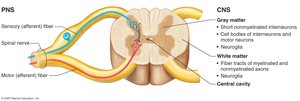
Composed of neuron cell bodies, dendrites, nonmyelinated axons, and neuroglia.
Forms the butterfly-shaped region in the spinal cord and surrounds central cavities.
Site of synapses and integration.
White Matter
Composed of myelinated axons (tracts) that connect different CNS regions.
Lies external to gray matter in the CNS.
Gross Anatomy of Nerves
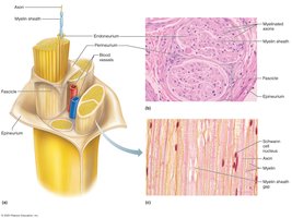
Nerves are cablelike organs in the PNS, consisting of bundles of axons (nerve fibers) wrapped in connective tissue layers:
Endoneurium: Surrounds individual axons.
Perineurium: Surrounds bundles of axons (fascicles).
Epineurium: Surrounds the entire nerve.
Neuronal Integration and Reflex Arcs
Neuronal Integration
The CNS and PNS are functionally interconnected. Sensory information is relayed to the CNS, which processes it and sends motor commands to effectors.
Reflex Arcs
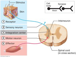
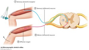
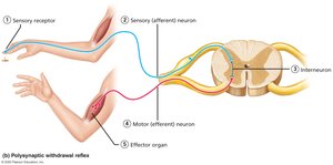
Reflex arcs are the basic structural and functional units of the nervous system, responsible for rapid, automatic responses to stimuli. They consist of five components:
Receptor: Detects the stimulus.
Sensory Neuron: Transmits afferent impulses to the CNS.
Integration Center: One or more synapses in the CNS.
Motor Neuron: Conducts efferent impulses to an effector.
Effector: Muscle or gland that responds to the stimulus.
Types of Reflexes
Monosynaptic Reflex: Involves a single synapse (e.g., knee-jerk reflex); fastest type.
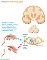
Polysynaptic Reflex: Involves one or more interneurons; more common (e.g., withdrawal reflex).
Integration Between PNS and CNS
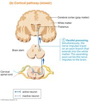
Neuronal circuits form complex networks that process sensory input and coordinate motor output. For example, a painful stimulus triggers an immediate spinal reflex and a slower, conscious perception of pain in the brain.
Disorders and Regeneration in the Nervous System
Multiple Sclerosis (MS)
An autoimmune disease where the immune system attacks myelin in the CNS.
Characterized by periods of relapse and remission; symptoms and severity vary widely.
Cause is not fully understood.
Neuronal Regeneration
PNS: If only axons are damaged, Schwann cells and macrophages can facilitate partial regeneration.
CNS: Regeneration is limited due to inhibitory factors produced by neuroglia; injuries often result in permanent dysfunction.