 뒤로
Chapter 11: Fundamentals of the Nervous System and Nervous Tissue: Structured Study Notes
스터디 가이드 - 스마트 노트
 자료에 맞춘 맞춤형 노트, 핵심 정의, 예시, 맥락을 확장해 제공합니다.
Fundamentals of the Nervous System and Nervous Tissue
The Nervous System: Overview and Functions
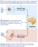
The nervous system is the master controlling and communicating system of the body. It uses electrical and chemical signals to rapidly and specifically coordinate responses to internal and external stimuli.
Sensory Input: Information is gathered by sensory receptors about internal and external changes.
Integration: Processing and interpretation of sensory input, deciding on a response.
Motor Output: Activation of effectors (muscles and glands) produces a response.
Example: Touching a hot object triggers sensory input, integration in the brain, and motor output to withdraw the hand.
Organization of the Nervous System
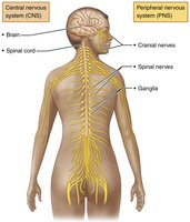
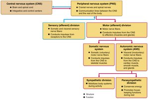
The nervous system is divided into two principal parts: the Central Nervous System (CNS) and the Peripheral Nervous System (PNS). Each has distinct structures and functions.
CNS: Consists of the brain and spinal cord; responsible for integration and control.
PNS: Consists mainly of nerves extending from the CNS; includes cranial and spinal nerves, and ganglia.
Functional Divisions of PNS:
Sensory (afferent) division: Carries information to the CNS.
Motor (efferent) division: Carries commands from the CNS to effectors.
Somatic Nervous System: Voluntary control of skeletal muscles.
Autonomic Nervous System (ANS): Involuntary control of smooth muscle, cardiac muscle, and glands; includes sympathetic and parasympathetic divisions.
Neuroglia and Neurons
Neuroglia (Glial Cells)
Neuroglia are supporting cells in nervous tissue. They surround and protect neurons, and are essential for proper nervous system function.
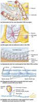
Astrocytes: Most abundant; support neurons, regulate environment, guide neuron migration, recycle neurotransmitters.
Microglial Cells: Monitor neurons, migrate to injury, phagocytize debris.
Ependymal Cells: Line CNS cavities, circulate cerebrospinal fluid (CSF).
Oligodendrocytes: Form myelin sheaths in CNS.
Satellite Cells (PNS): Surround neuron cell bodies, similar to astrocytes.
Schwann Cells (PNS): Form myelin sheaths, aid in nerve regeneration.
Neurons: Structure and Function
Neurons are excitable cells that transmit electrical signals. They have unique properties and structures that enable rapid communication.
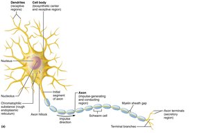
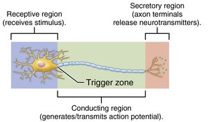
Cell Body (Soma): Contains nucleus, metabolic center, and organelles.
Dendrites: Receptive regions; convey signals toward cell body.
Axon: Conducting region; transmits impulses away from cell body to axon terminals.
Axon Terminals: Secretory region; release neurotransmitters.
Myelin Sheath: Insulates axons, increases impulse speed.
Classification of Neurons
Structural Classification
Neurons are classified by the number of processes extending from the cell body.
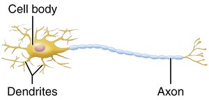
Multipolar: Many dendrites, one axon; most common in CNS.
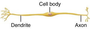
Bipolar: One dendrite, one axon; found in retina, olfactory mucosa.
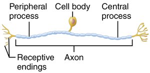
Unipolar: Single process divides into two branches; sensory neurons in PNS.
Functional Classification
Neurons are also classified by the direction of impulse conduction.
Sensory (afferent) neurons: Transmit impulses toward CNS.
Motor (efferent) neurons: Carry impulses from CNS to effectors.
Interneurons: Connect sensory and motor neurons; most abundant in CNS.
Membrane Potentials and Electrical Properties
Basic Principles of Electricity
Neurons use electrical signals to communicate. Key concepts include voltage, current, and resistance, governed by Ohm's law:
Voltage (V): Potential energy from separated charges.
Current (I): Flow of electrical charge (ions).
Resistance (R): Hindrance to charge flow.
Ohm's Law:
Resting Membrane Potential
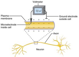
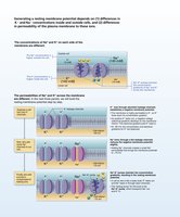
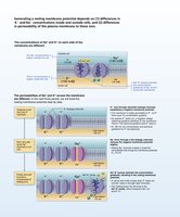
The resting membrane potential is the voltage across the neuron's membrane when not transmitting signals, typically around -70 mV. It is established by differences in ion concentrations and membrane permeability.
Key ions: Sodium (Na+), Potassium (K+), Chloride (Cl-).
Sodium-potassium pump: Maintains gradients by pumping 3 Na+ out and 2 K+ in.
Polarization: Inside of cell is more negative than outside.
Changes in Membrane Potential
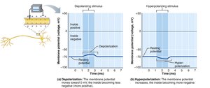
Membrane potential changes are essential for neural signaling. Depolarization decreases membrane potential, while hyperpolarization increases it.
Depolarization: Membrane potential moves toward zero; increases likelihood of action potential.
Hyperpolarization: Membrane potential becomes more negative; decreases likelihood of action potential.
Graded Potentials and Action Potentials
Graded Potentials
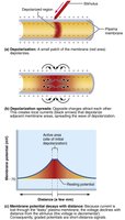
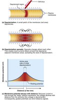
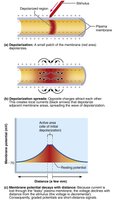
Graded potentials are short-lived, localized changes in membrane potential. They act as signals over short distances and are essential for initiating action potentials.
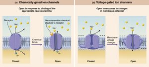
Triggered by: Opening of gated ion channels.
Types: Receptor potential, postsynaptic potential, end-plate potential.
Spread and Decay: Current flows but dissipates quickly.
Action Potentials
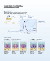
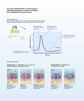
An action potential (AP) is a brief reversal of membrane potential, used for long-distance signaling in neurons and muscle cells. APs do not decay over distance.
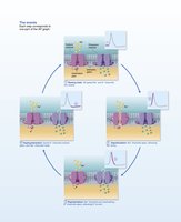
Four main steps:
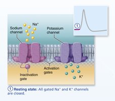
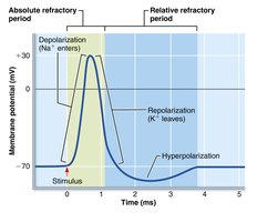
Resting state: All voltage-gated channels closed.
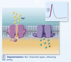
Depolarization: Na+ channels open, Na+ enters.
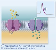
Repolarization: Na+ channels inactivate, K+ channels open, K+ exits.
Hyperpolarization: Some K+ channels remain open, membrane becomes more negative.
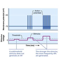
Threshold: Minimum depolarization required to trigger AP.
All-or-none phenomenon: AP either occurs fully or not at all.
Propagation and Coding of Action Potentials
Action potentials propagate along axons, allowing rapid transmission of signals. The frequency of APs encodes stimulus intensity.
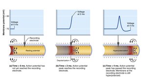
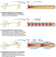
Propagation: AP moves from origin down axon; self-propagating.
Conduction velocity: Depends on axon diameter and myelination.
Saltatory conduction: AP jumps between nodes of Ranvier in myelinated axons; much faster than continuous conduction in unmyelinated axons.
Stimulus intensity: Higher frequency of APs indicates stronger stimulus.
Refractory Periods
Refractory periods ensure unidirectional propagation and limit AP frequency.
Absolute refractory period: No new AP can be generated.
Relative refractory period: AP can be generated only by a strong stimulus.
Conduction Velocity
Conduction velocity varies based on axon diameter and myelination.
A fibers: Largest diameter, heavily myelinated, fastest (150 m/s).
B fibers: Intermediate diameter, lightly myelinated.
C fibers: Smallest diameter, nonmyelinated, slowest.
Synapses and Neurotransmitters
Synapses
Synapses are junctions that mediate information transfer between neurons or from neurons to effector cells. There are electrical and chemical synapses.
Presynaptic neuron: Sends information.
Postsynaptic neuron: Receives information.
Types: Axodendritic, axosomatic, axoaxonal, dendrodendritic, somatodendritic.
Electrical synapses: Direct, rapid communication via gap junctions.
Chemical synapses: Use neurotransmitters; most common.
Chemical Synapse Transmission
Transmission across chemical synapses involves six steps:
AP arrives at axon terminal.
Voltage-gated Ca2+ channels open; Ca2+ enters.
Ca2+ entry causes synaptic vesicles to release neurotransmitter by exocytosis.
Neurotransmitter diffuses across synaptic cleft and binds to receptors on postsynaptic membrane.
Binding opens ion channels, creating graded potentials.
Neurotransmitter effects are terminated by reuptake, degradation, or diffusion.
Postsynaptic Potentials and Summation
Excitatory and Inhibitory Postsynaptic Potentials
Neurotransmitter binding causes graded potentials in the postsynaptic neuron.
EPSP (Excitatory): Depolarizes membrane, increases likelihood of AP.
IPSP (Inhibitory): Hyperpolarizes membrane, decreases likelihood of AP.
Summation
Multiple EPSPs and IPSPs can summate to influence whether an AP is generated.
Temporal summation: Rapid-fire impulses from one neuron.
Spatial summation: Simultaneous impulses from multiple neurons.
Neurotransmitter Classification and Function
Chemical Structure
Acetylcholine: Released at neuromuscular junctions.
Biogenic amines: Dopamine, norepinephrine, serotonin, histamine.
Amino acids: Glutamate, aspartate, glycine, GABA.
Peptides: Substance P, endorphins.
Purines: ATP, adenosine.
Gases and lipids: Nitric oxide, carbon monoxide, endocannabinoids.
Functional Classification
Excitatory vs. Inhibitory: Effect depends on receptor type.
Direct vs. Indirect: Direct action opens ion channels; indirect action uses second messengers.
Neuromodulators: Modify synaptic transmission strength.
Neural Integration and Circuits
Patterns of Neural Processing
Serial processing: One pathway, specific response (e.g., reflex arc).
Parallel processing: Multiple pathways, complex responses.
Types of Neural Circuits
Diverging: One input, many outputs.
Converging: Many inputs, one output.
Reverberating: Repetitive signals.
Parallel after-discharge: Multiple pathways, single output.
Developmental Aspects of Neurons
The nervous system originates from the neural tube and neural crest. Neurons undergo proliferation, migration, and connection. Synaptic pruning and apoptosis shape the mature nervous system.
Growth cone: Guides axon to target.
Astrocytes: Support synapse formation.
Apoptosis: Programmed cell death eliminates excess neurons.
Additional info: These notes are based on Chapter 11 of Marieb Human Anatomy & Physiology, covering the fundamentals of the nervous system and nervous tissue, including structure, function, electrical properties, synaptic transmission, and neural integration.
