 뒤로
Heart and Cardiovascular Physiology: Structured Study Notes
스터디 가이드 - 스마트 노트
 자료에 맞춘 맞춤형 노트, 핵심 정의, 예시, 맥락을 확장해 제공합니다.
Heart Anatomy
Location, Structure, and Blood Supply
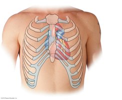
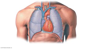
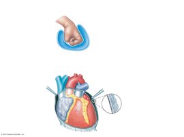
The heart is a muscular organ located in the mediastinum of the thoracic cavity, directly deep to the sternum. It is roughly the size of a fist, with its base tilted toward the right shoulder and its apex pointing toward the left hip. The mediastinum also contains the esophagus, trachea, thymus, and major blood vessels.
Base: The broad, flat upper part of the heart.
Apex: The pointed lower part of the heart.
Mediastinum: Central compartment of the thoracic cavity.
Layers of the Heart
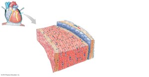
The heart wall consists of three main layers, each with distinct structure and function. Surrounding the heart is the pericardium, a double-layered sac that protects and anchors the heart.
Endocardium: Inner lining made of simple squamous epithelium and areolar connective tissue.
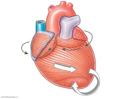
Myocardium: Middle layer composed of cardiac muscle tissue, responsible for contraction.
Epicardium (Visceral Pericardium): Outer layer, part of the serous membrane.
Parietal Pericardium: Outer serous layer, reinforced by fibrous pericardium.
Pericardial Cavity: Space between serous layers, filled with serous fluid to reduce friction.
Cardiac Muscle Tissue
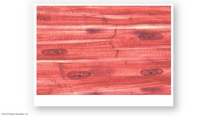
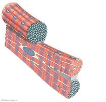
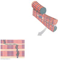
Cardiac muscle cells are smaller than skeletal muscle fibers, striated, and typically have a single, centrally located nucleus. They rely heavily on aerobic respiration, containing numerous mitochondria and high levels of myoglobin. Cardiac muscle cells branch and connect via intercalated discs, which facilitate synchronized contraction.
Intercalated Discs: Specialized junctions that allow direct electrical and chemical communication between cells.
Gap Junctions: Permit ions and small molecules to pass, enabling action potentials to spread rapidly.
Desmosomes: Provide mechanical strength and anchoring support.
External Features of the Heart
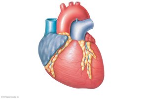
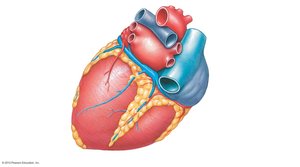
The heart has four chambers: right and left atria (upper chambers) and right and left ventricles (lower chambers). Sulci (grooves) mark the boundaries between chambers and contain blood vessels and adipose tissue. The coronary sulcus separates atria from ventricles, while the anterior and posterior interventricular sulci divide the right and left ventricles. Major vessels include the superior and inferior vena cava, pulmonary trunk, pulmonary veins, and aorta.
Heart’s Blood Supply
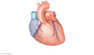
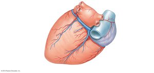
The myocardium receives blood from the right and left coronary arteries, which branch from the ascending aorta. Cardiac veins drain the myocardium and converge in the coronary sinus, which empties into the right atrium.
Coronary Arteries: Supply oxygenated blood to the heart muscle.
Cardiac Veins: Remove deoxygenated blood from the myocardium.
Coronary Sinus: Main venous drainage into the right atrium.
Chambers, Valves, and Great Vessels
Heart Chambers and Circulation
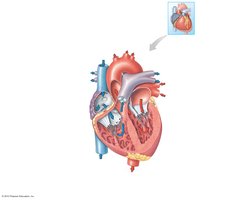
The heart is divided into four chambers, each with specific roles in circulation. The right atrium receives deoxygenated blood from the body, the right ventricle pumps it to the lungs, the left atrium receives oxygenated blood from the lungs, and the left ventricle pumps it to the body.
Atria: Receiving chambers; pump blood into ventricles.
Ventricles: Pump blood out to pulmonary and systemic circuits.
Interatrial Septum: Wall between atria.
Interventricular Septum: Wall between ventricles.
Fossa Ovalis: Remnant of fetal foramen ovale.
Heart Valves
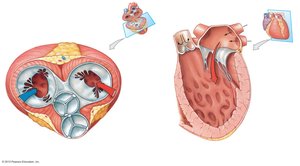
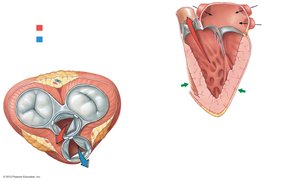
Valves ensure unidirectional blood flow through the heart. Atrioventricular (AV) valves allow blood flow from atria to ventricles, while semilunar (SL) valves allow blood flow from ventricles to arteries.
Tricuspid Valve: Right AV valve.
Bicuspid (Mitral) Valve: Left AV valve.
Pulmonary Valve: SL valve between right ventricle and pulmonary trunk.
Aortic Valve: SL valve between left ventricle and aorta.
Chordae Tendineae: Fibrous strings attaching valve cusps to papillary muscles.
Papillary Muscles: Contract to prevent valve prolapse.
Great Vessels
Major vessels associated with the heart include the superior and inferior vena cava, pulmonary trunk and arteries, pulmonary veins, and aorta. These vessels are essential for systemic and pulmonary circulation.
Superior/Inferior Vena Cava: Return deoxygenated blood to right atrium.
Pulmonary Trunk/Arteries: Carry deoxygenated blood to lungs.
Pulmonary Veins: Return oxygenated blood to left atrium.
Aorta: Distributes oxygenated blood to body tissues.
Route of Blood Flow Through the Heart
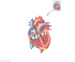
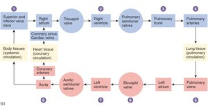
Blood flows through the heart in a specific sequence, passing through chambers and valves to ensure proper oxygenation and circulation.
Deoxygenated blood enters right atrium → right ventricle → pulmonary arteries → lungs.
Oxygenated blood returns via pulmonary veins → left atrium → left ventricle → aorta → body.
Cardiac Cycle and Cardiac Conduction
Cardiac Cycle
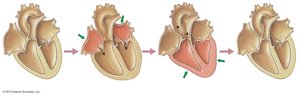
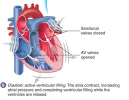
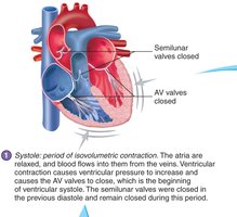
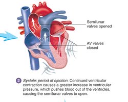
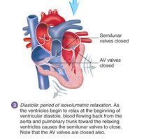
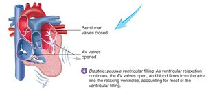
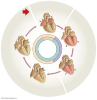
The cardiac cycle consists of alternating periods of contraction (systole) and relaxation (diastole) in the heart chambers. Both sides of the heart contract and relax simultaneously, ensuring efficient blood flow.
Systole: Contraction phase; blood is ejected.
Diastole: Relaxation phase; chambers fill with blood.
Heart Sounds
Heart sounds are produced by turbulent blood flow during valve closure. The first sound (S1, "lubb") is due to AV valve closure; the second sound (S2, "dupp") is due to SL valve closure. S3 and S4 are rarely heard in adults.

Electrical Conduction
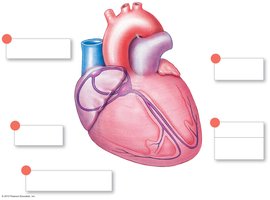
The heart's electrical system initiates and coordinates contraction. The sinoatrial (SA) node acts as the pacemaker, generating action potentials that spread through the atria and reach the atrioventricular (AV) node. The AV bundle, bundle branches, and Purkinje fibers distribute the signal to the ventricles.
SA Node: Pacemaker; initiates heartbeat.
AV Node: Delays and relays signal to ventricles.
Purkinje Fibers: Rapidly conduct signal to ventricular myocardium.
Regulation of Heart Rate
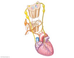
Heart rate is regulated by autonomic nervous system signals and hormones. Parasympathetic stimulation (via vagus nerve) slows the heart, while sympathetic stimulation (via cardiac nerves and hormones) increases heart rate.
Bradycardia: Slow heart rate (<60 bpm).
Tachycardia: Fast heart rate (>100 bpm).
Electrocardiogram (ECG/EKG)
An ECG records the electrical activity of the heart. The P wave represents atrial depolarization, the QRS complex represents ventricular depolarization, and the T wave represents ventricular repolarization. Arrhythmias are detected as irregular patterns.
P Wave: Atrial depolarization.
QRS Complex: Ventricular depolarization.
T Wave: Ventricular repolarization.
Cardiac Output, Blood Flow, and Capillary Exchange
Cardiac Output
Cardiac output (CO) is the volume of blood ejected by the heart per minute. It is calculated as the product of heart rate (HR) and stroke volume (SV).
Formula:
Example: If HR = 75 beats/min and SV = 80 mL/beat, then mL/min.
Heart Failure: Occurs when CO cannot meet tissue demands.

Factors Affecting Cardiac Output
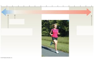
Cardiac output is influenced by factors affecting stroke volume and heart rate, including neural and hormonal regulation, venous return, contractility, and body temperature.
Neural Regulation: Autonomic nervous system control.
Hormonal Regulation: Epinephrine, norepinephrine, and other hormones.
Venous Return: Amount of blood returning to the heart.
Contractility: Force of myocardial contraction.
Blood Flow
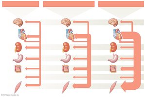
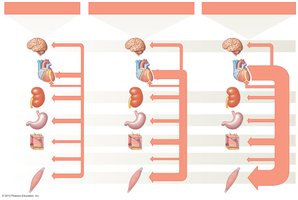
Blood flow is the volume of blood moving past a point per unit time. It is directly proportional to blood pressure and inversely proportional to peripheral resistance, which is mainly determined by vessel diameter.
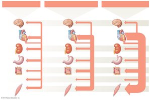
During Exercise: Blood flow distribution shifts to prioritize skeletal muscles.
Capillary Exchange
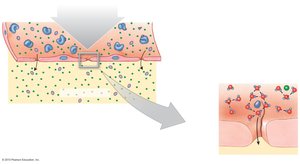
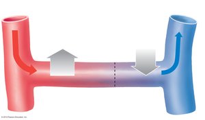
Capillary exchange involves filtration, diffusion, and osmosis. Filtration occurs when plasma and solutes are forced out of capillaries by hydrostatic pressure. Net filtration pressure is the difference between hydrostatic and osmotic pressures.
Filtration: Movement of fluid out of capillaries.
Diffusion: Movement of molecules between blood and tissues.
Osmosis: Water movement driven by osmotic pressure.
Formula:
Hydrostatic Pressure:
Osmotic Pressure:
Blood Pressure Regulation
Blood Pressure
Blood pressure is the force exerted by blood within the cardiovascular system. It is highest in arteries and lowest in veins. Pulse pressure is the difference between systolic and diastolic pressures in arteries.
High BP: Can damage vessels.
Low BP: Reduces blood flow to tissues.
Autoregulation and Central Regulation
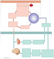
Autoregulation involves local control of blood flow via precapillary sphincters, responding to chemical and metabolic changes. Central regulation includes neural (short-term) and endocrine (long-term) mechanisms to maintain homeostasis.
Neural Mechanisms: Adjust heart rate and vessel diameter.
Endocrine Mechanisms: Influence blood volume and pressure.
Baroreceptor Reflex
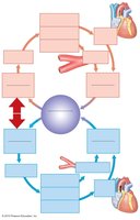
The baroreceptor reflex is a key short-term regulator of blood pressure. Baroreceptors in the carotid body and aortic arch detect changes in pressure and trigger vasodilation or vasoconstriction as needed.
High BP: Vasodilation and decreased cardiac output.
Low BP: Vasoconstriction and increased cardiac output.
Endocrine Regulation
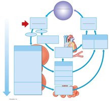
Endocrine responses to blood pressure changes include the renin-angiotensin-aldosterone system and antidiuretic hormone (ADH) release. The kidneys release renin when blood pressure drops, leading to angiotensin II formation, which causes vasoconstriction and stimulates ADH and aldosterone release.
Renin-Angiotensin-Aldosterone System: Increases blood pressure and volume.
ADH: Promotes water retention.
EPO: Stimulates red blood cell production if oxygen is low.
Atrial Natriuretic Peptide (ANP)
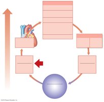
ANP is a hormone released by the heart when atrial stretch is excessive. It promotes sodium and water loss, reduces thirst, inhibits ADH and aldosterone, and causes peripheral vasodilation, lowering blood pressure and volume.
ANP Effects: Increased Na+ and water loss, reduced blood volume and pressure.
 Additional info: These notes provide a comprehensive overview of heart anatomy, physiology, and regulatory mechanisms, suitable for college-level Anatomy & Physiology students.
