뒤로
Heart and Cardiovascular Physiology: Structured Study Notes
스터디 가이드 - 스마트 노트
 자료에 맞춘 맞춤형 노트, 핵심 정의, 예시, 맥락을 확장해 제공합니다.
Heart Anatomy
Location, Structure, and Blood Supply
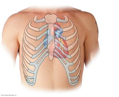
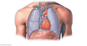
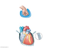
The heart is a muscular organ located in the mediastinum of the thoracic cavity, directly deep to the sternum. It is about the size of a fist, with its base tilted toward the right shoulder and its apex toward the left hip. The mediastinum also contains the esophagus, trachea, thymus, and major blood vessels.
Base: The broad, flat part of the heart, oriented toward the right shoulder.
Apex: The pointed end, oriented toward the left hip.
Mediastinum: Central compartment of the thoracic cavity.
Layers of the Heart
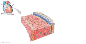
The heart wall consists of three main layers, each with distinct structure and function:
Endocardium: Inner lining composed of simple squamous epithelium and areolar connective tissue.
Myocardium: Middle layer made of cardiac muscle tissue, responsible for contraction. Contains blood vessels and nerves.
Epicardium (Visceral Pericardium): Outer layer, part of the serous membrane surrounding the heart.
Parietal Pericardium: Parietal serous layer, reinforced by fibrous pericardium.
Pericardial Cavity: Space between serous layers, filled with serous fluid to reduce friction.
Cardiac Muscle Tissue
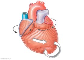
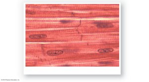
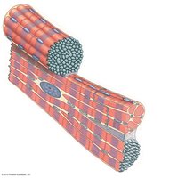
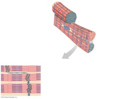
Cardiac muscle cells are smaller than skeletal muscle fibers, striated, and typically have a single, centrally located nucleus. They rely heavily on aerobic respiration, containing many mitochondria and high levels of myoglobin. Cardiac muscle cells branch and connect via intercalated discs, which contain gap junctions and desmosomes for electrical and mechanical connectivity.
Intercalated Discs: Specialized junctions that allow direct electrical communication and anchoring between cells.
Gap Junctions: Permit ion flow for synchronized contraction.
Desmosomes: Provide mechanical strength.
External Features of the Heart
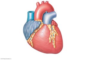
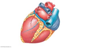
The heart has four chambers: right and left atria (upper chambers) and right and left ventricles (lower chambers). Sulci (grooves) mark divisions between chambers and contain adipose tissue and blood vessels. The coronary sulcus divides atria from ventricles, while anterior and posterior interventricular sulci divide the ventricles. Major vessels include the superior and inferior vena cava, pulmonary trunk, pulmonary veins, and aorta.
Heart’s Blood Supply
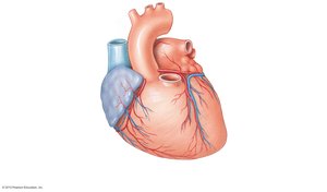
The right and left coronary arteries are the first branches from the ascending aorta, supplying the myocardium. Cardiac veins drain the myocardium and converge in the coronary sinus, which empties into the right atrium.
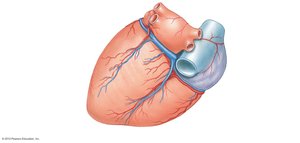
Chambers, Valves, and Great Vessels
Heart Chambers and Circulation
The heart is divided into four chambers, each with specific roles in circulation:
Right Atrium: Receives deoxygenated blood from the systemic circuit.
Right Ventricle: Pumps deoxygenated blood into the pulmonary circuit.
Left Atrium: Receives oxygenated blood from the pulmonary circuit.
Left Ventricle: Pumps oxygenated blood into the systemic circuit.
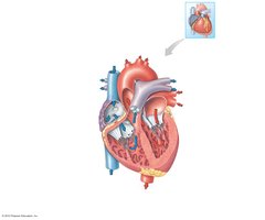
Features of the Heart Chambers
Atria: Receiving chambers; pump blood down to ventricles.
Auricles: Flaps on atria for extra volume.
Pectinate Muscles: Ridges in atria and auricles.
Ventricles: Pump blood out to pulmonary and systemic circuits.
Trabeculae Carneae: Columns and ridges in ventricles.
Interatrial Septum: Wall between atria.
Fossa Ovalis: Remnant of fetal foramen ovale.
Interventricular Septum: Wall between ventricles.
Heart Valves
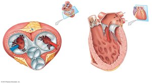
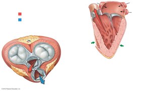
Valves ensure one-way flow of blood:
Atrioventricular (AV) Valves: Tricuspid (right) and bicuspid/mitral (left); allow passage from atrium to ventricle.
Semilunar (SL) Valves: Pulmonary (right) and aortic (left); allow passage from ventricle to artery.
Chordae Tendineae: Attach valve cusps to papillary muscles.
Papillary Muscles: Contract to prevent valve prolapse.
Great Vessels
Superior Vena Cava: Returns deoxygenated blood from above diaphragm.
Inferior Vena Cava: Returns deoxygenated blood from below diaphragm.
Pulmonary Trunk: Carries deoxygenated blood to lungs.
Pulmonary Veins: Return oxygenated blood from lungs.
Aorta: Distributes oxygenated blood to body.
Ligamentum Arteriosum: Remnant of fetal ductus arteriosus.
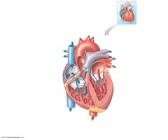
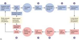
Route of Blood Flow Through Heart
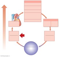
Blood flows through the heart in a specific sequence, ensuring oxygenation and distribution to tissues.
Cardiac Cycle and Cardiac Conduction
Cardiac Cycle
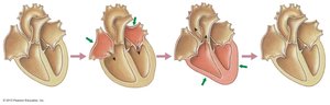
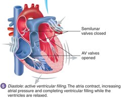
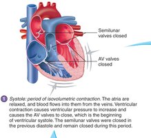
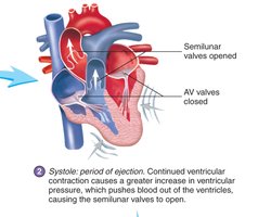
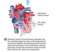
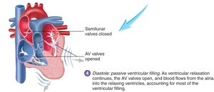
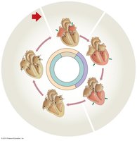
The cardiac cycle consists of alternating periods of contraction (systole) and relaxation (diastole) in the atria and ventricles. Both sides of the heart work simultaneously, with atria contracting together and ventricles contracting together.
Systole: Contraction phase.
Diastole: Relaxation phase.
Heart Sounds
Heart sounds are produced by turbulent blood flow during valve closure:
First Heart Sound (S1, lubb): Closing of AV valves at start of ventricular systole.
Second Heart Sound (S2, dupp): Closing of SL valves at start of ventricular diastole.
Third and Fourth Sounds: Usually not heard in adults.

Electrical Conduction
The heart's electrical system coordinates contraction:
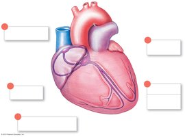
Sinoatrial (SA) Node: Pacemaker in right atrium.
Internodal Pathways: Spread signal across atria.
Atrioventricular (AV) Node: Delays signal, sends to ventricles.
AV Bundle and Bundle Branches: Conduct signal down septum.
Purkinje Fibers: Spread signal through ventricles.
Regulation of Heart Rate
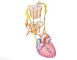
Heart rate is regulated by autonomic nervous system and hormones:
Parasympathetic: Vagus nerve slows heart rate.
Sympathetic: Cardiac nerves and hormones (epinephrine, norepinephrine) increase heart rate.
Bradycardia: Slow heart rate (<60 bpm).
Tachycardia: Fast heart rate (>100 bpm).
Electrocardiogram (ECG/EKG)
An ECG records the electrical activity of the heart:
P Wave: Atrial depolarization.
QRS Complex: Ventricular depolarization.
T Wave: Ventricular repolarization.
Cardiac Output, Blood Flow, and Capillary Exchange
Cardiac Output
Cardiac output (CO) is the volume of blood ejected by the heart per minute. It is calculated as:
CO = Heart Rate (HR) × Stroke Volume (SV)
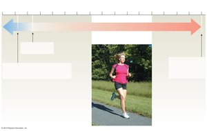
During exercise, CO increases significantly. Heart failure occurs when CO cannot meet tissue demands.

Factors Affecting Cardiac Output
Stroke Volume: Influenced by contractility, venous return, and hormonal/neural regulation.
Heart Rate: Influenced by neural and hormonal regulation, body temperature.
Blood Flow
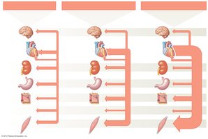
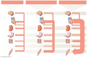
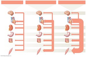
Blood flow is the volume of blood moving past a point per unit time. It is directly proportional to blood pressure and inversely proportional to peripheral resistance, which is mainly controlled by vessel diameter.
During exercise: Blood flow distribution shifts to prioritize skeletal muscles.
Capillary Exchange
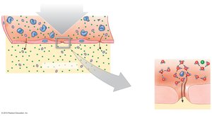
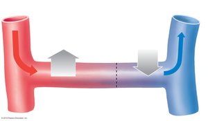
Capillary exchange involves filtration, diffusion, and osmosis:
Filtration: Plasma and solutes move out due to hydrostatic pressure.
Diffusion: Molecules move down concentration gradients.
Osmosis: Water moves in response to osmotic pressure.
Net filtration pressure is calculated as:
Net Filtration Pressure = Net Hydrostatic Pressure – Net Osmotic Pressure
NHP = Capillary Hydrostatic Pressure – Interstitial Fluid Hydrostatic Pressure
NOP = Blood Colloid Osmotic Pressure – Interstitial Fluid Colloid Osmotic Pressure
Blood Pressure Regulation
Blood Pressure
Blood pressure is the force exerted by blood within the cardiovascular system. It is highest in arteries and lowest in veins. Pulse pressure is the difference between systolic and diastolic pressures in arteries.
Low BP: Reduces blood flow.
High BP: Damages vessels.
Autoregulation and Central Regulation
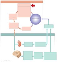
Blood pressure is regulated locally (autoregulation) and systemically (central regulation):
Autoregulation: Local vasodilators adjust blood flow based on tissue needs.
Central Regulation: Neural and endocrine mechanisms adjust blood pressure and volume.
Baroreceptor Reflex
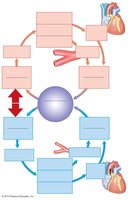
Baroreceptors in the carotid body and aortic arch detect changes in blood pressure and trigger reflexes:
High BP: Causes vasodilation and decreased cardiac output.
Low BP: Causes vasoconstriction and increased cardiac output.
Endocrine Regulation
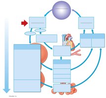
Hormones play a key role in long-term blood pressure regulation:
Renin-Angiotensin-Aldosterone System: Increases blood pressure and volume via vasoconstriction and sodium/water retention.
Antidiuretic Hormone (ADH): Promotes water retention.
Erythropoietin (EPO): Stimulates red blood cell production.
Atrial Natriuretic Peptide (ANP)
ANP is released by the heart when atria are stretched, promoting sodium and water loss, reducing blood volume and pressure.